

Stereotactic Radiosurgery QA: MRI/CT Fusion
In stereotactic radiosurgery, a planning image is only useful when its geometry can be trusted. MRI can add valuable soft-tissue information, but distortion and registration differences may affect how anatomy corresponds with CT-based planning data. That makes imaging verification a distinct part of the broader QA process.
Stereotactic radiosurgery QA can use an MRI/CT fusion phantom to evaluate geometric distortion, marker-based co-registration, and spatial agreement within the relevant scan field of view. These tests help teams document imaging and registration behavior; they do not replace end-to-end treatment-delivery QA or establish clinical safety.
For medical physicists and treatment-planning teams, the practical question is how to measure these behaviors consistently and interpret the results within a site-specific workflow. That begins with understanding why even small geometric discrepancies matter when imaging data guides stereotactic planning.
Why geometric distortion matters in SRS treatment planning
Stereotactic radiosurgery QA is the structured process of evaluating the imaging, planning, positioning, and delivery steps that support highly precise radiation treatment. Stereotactic planning depends on coordinate systems, immobilization, and diagnostic imaging. So the spatial relationship between anatomy and treatment-planning coordinates must be examined as part of a local quality-assurance program. A useful overview of radiation therapy QA phantoms can help place this imaging work within the broader planning and delivery workflow.
MRI geometric distortion matters because the anatomy represented in an MR image may not correspond perfectly to its physical position. Distortion can vary across the head scan field of view and may be influenced by the scanner, sequence, gradients, susceptibility effects, and acquisition setup. When MRI is registered to CT for treatment planning, any spatial discrepancy can affect the correspondence between image sets. The registration algorithm may also introduce behavior that needs to be evaluated rather than assumed.
This is especially important when the planning process uses small spatial margins. The planning target volume is intended to account for systematic and random uncertainties in treatment planning and dose delivery. A review of SRS and stereotactic radiotherapy describes 1 mm translational and 1 degree rotational shifts as a general principle. But those values should not be treated as a universal acceptance criterion for every institution or workflow. Local protocols, equipment, immobilization, registration methods, and clinical risk assessments determine how results are interpreted.
What imaging QA can show
An imaging-focused evaluation can help a medical physicist quantify geometric behavior, inspect MRI-to-CT correspondence, and document whether a registration workflow behaves consistently under defined conditions. For example, a phantom with known spatial features can provide reference points for reviewing distortion across the head field of view. MRI/CT-compatible markers can support co-registration alignment, while a structured analysis can separate scanner-related image geometry from software registration effects.
Radiology Support Devices Inc., trading as RSD Phantoms, documents the Multi-Modality Fusion Head Phantom as a tool for MRI/CT fusion verification and geometric distortion measurements. Its documented scope includes evaluating image fusion, registration software, image quality, and protocol verification. These are imaging and planning-system checks, not claims about patient diagnosis, treatment, clinical safety, or dose-delivery accuracy.
Why imaging QA does not replace delivery QA
Imaging QA and end-to-end treatment delivery QA answer different questions. Imaging QA asks whether the acquired images and registration process preserve the spatial information needed for planning. End-to-end QA follows a broader chain, which may include target localization, treatment-plan calculation, patient-specific setup, beam delivery, and measurement of the delivered result. A fusion phantom can help investigate image distortion and registration, but it should not be presented as proof that the complete treatment-delivery process is accurate. Keeping these boundaries explicit makes the QA record more useful and helps the team select complementary tests for each stage of the SRS workflow.
How MRI/CT fusion phantoms evaluate spatial accuracy
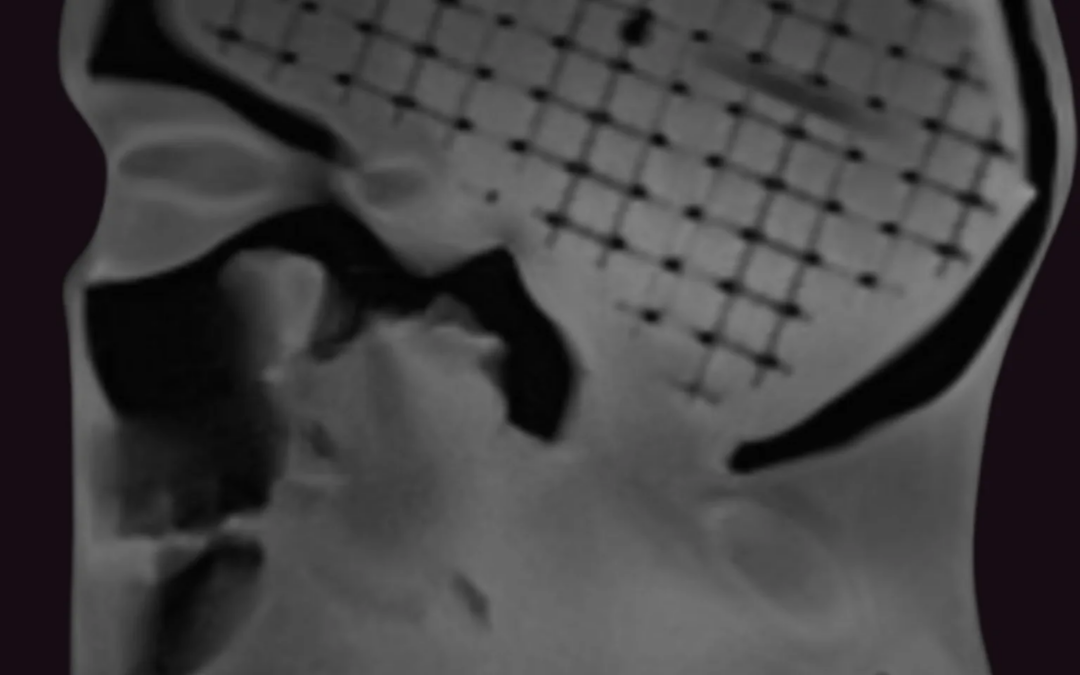
A practical MRI/CT fusion QA workflow compares the same physical geometry across image sets, then examines whether registration software preserves that geometry. This makes the test more specific than reviewing image quality in either modality alone. For stereotactic radiosurgery QA, the goal is to identify where distortion, marker localization, or registration behavior could affect spatial correspondence during treatment planning. The MRI/CT fusion QA phantom is designed for this type of MRI/CT fusion validation using geometric distortion measurements.
- Plan the scan and evaluation. Define the MRI sequences, CT acquisition, head-coil arrangement, field of view, slice geometry, reconstruction settings, and treatment-planning or registration software to be evaluated. Keep the intended QA boundary explicit. This workflow examines imaging geometry, fusion, and registration behavior. It does not by itself establish dose-delivery accuracy or provide a clinical safety guarantee. Confirm that the selected configuration is appropriate for the scanners and software in use rather than assuming universal compatibility.
- Position the phantom and acquire reference images. Place the phantom consistently and document the setup so the test can be repeated after a protocol, coil, scanner, or software change. Acquire the MRI and CT data using the protocols under review. The Multi-Modality Fusion Head Phantom includes MRI/CT-compatible markers that support co-registration alignment. Its documented design also includes a proprietary aqueous solution that generates soft-tissue signals for T1-, T2-, and proton-density-weighted imaging. This allows the team to assess the behavior of the selected MRI protocols within the intended use case.
- Inspect the lattice and distortion pattern. Review the three-dimensional lattice across the head scan field of view. The lattice is designed to evaluate MR distortion, so the reviewer can inspect whether the imaged geometry remains spatially consistent across the region relevant to planning. Record the scan protocol, field-of-view coverage, observed distortion pattern, and analysis method. Avoid assigning a universal pass or fail threshold unless one has been established by the local program, protocol, or applicable documentation.
- Perform marker-based co-registration. Load the MRI and CT datasets into the treatment-planning or image-registration environment and use the compatible markers to establish the initial alignment. Compare the resulting correspondence against the known phantom geometry. The purpose is to evaluate how the registration workflow handles the relationship between MRI and CT, not simply whether the images appear visually similar in a review window.
- Assess fusion and deformable-registration behavior. Evaluate the image-fusion result throughout the relevant field of view, including areas where MR distortion may influence spatial correspondence. If deformable image-registration software is part of the clinical workflow, test its behavior separately from rigid co-registration and document which settings were used. The product is described as a verification phantom for image fusion and deformable image-registration software in treatment-planning systems. So findings should remain tied to the tested configuration and workflow.
- Document findings and define follow-up. Preserve the datasets, acquisition parameters, registration settings, visual findings, measurements, reviewer identity, and disposition of any discrepancy. Compare results with the program’s established baseline or acceptance process. The product documentation identifies MRI/CT fusion verification, MRI geometric-distortion measurement, image quality, and protocol verification as intended applications. Use those results to determine whether a protocol needs investigation or repeat testing, while keeping imaging QA distinct from separate end-to-end treatment and dose-delivery checks.
How Stereotactic Radiosurgery QA Evaluates MRI/CT Fusion
A useful MRI geometric distortion QA workflow measures more than whether a single image appears acceptable. It should show how spatial fidelity behaves across the head scan field of view. It should also assess whether identifiable structures align between MRI and CT and whether the result remains consistent when the imaging protocol is repeated. For medical physicists evaluating MRI/CT fusion QA, the goal is to create a repeatable evidence trail for imaging and registration performance.
The Multi-Modality Fusion Head Phantom is documented for MRI/CT fusion verification, MRI geometric-distortion measurements, image quality, and protocol verification. Its 3D lattice is designed to evaluate MR distortion within the head field of view, while MRI/CT-compatible markers support co-registration alignment. The product documentation also describes a proprietary aqueous solution that generates soft-tissue signals for T1-, T2-, and proton-density-weighted imaging. These features support a structured assessment, but they do not establish a universal acceptance limit or a clinical safety guarantee.
| Measurement area | What to evaluate | Why it matters |
|---|---|---|
| Distortion across the head field of view | Review the 3D lattice throughout the head scan field of view, rather than checking only the central region. Compare measured spatial relationships across the area used by the planning workflow. | Geometric distortion may vary with position. A field-wide review helps identify where spatial representation changes and where additional local investigation may be appropriate. |
| Marker and co-registration alignment | Confirm that MRI/CT-compatible markers can be identified consistently and that the registered datasets preserve their expected correspondence. | Marker behavior provides a practical reference for reviewing alignment between modalities before assessing downstream registration behavior. |
| Image-fusion or registration behavior | Evaluate the fusion workflow and, where used, deformable image-registration software in the treatment-planning system. Record the registration method and observed spatial differences. | Registration performance is a separate question from scanner distortion. Testing both helps distinguish an acquisition issue from a fusion or software-workflow issue. |
| Protocol repeatability | Repeat the defined acquisition and analysis under the same documented conditions, including the relevant sequences and setup. Compare results across runs. | Repeatability shows whether observed differences are stable features of the workflow or variation introduced by setup, acquisition, or analysis. |
| Documentation | Record scanner and coil information, sequence parameters, phantom setup, field of view, registration method, analysis approach, observations, and disposition of findings. | A complete record makes the assessment reproducible and supports comparison after protocol, hardware, or software changes. |
This framework is intentionally measurement-focused. The article does not establish a universal pass/fail threshold for distortion, registration, or repeatability. Acceptance criteria should be defined by the local imaging and treatment-planning program, its documented protocols, and its risk assessment. Phantom testing can evaluate image distortion, fusion, registration, and protocol verification. It should not be presented as a substitute for end-to-end treatment-delivery QA, nor as evidence of dose-delivery accuracy or clinical safety.
How to interpret tolerance thresholds without overclaiming
A tolerance threshold is a decision point within a defined QA protocol, not a universal property of every stereotactic radiosurgery workflow. The planning target volume is used to account for systematic and random uncertainties in treatment planning and dose delivery. The appropriate action limit still depends on how a particular program measures, reports, and manages those uncertainties. A threshold should therefore be read alongside the imaging system, anatomy, registration method, immobilization approach, and clinical risk assessment.
A recent review describes 1 mm for translational shifts and 1 degree for rotational shifts as a general principle for SRS and stereotactic radiotherapy. That statement is useful as technical context, but it should not be presented as a universal acceptance criterion for every scanner, treatment-planning system, phantom, or patient-specific workflow. The review’s wording does not replace local commissioning documentation or an approved departmental tolerance policy. Teams should document whether the value is an investigation threshold, an action limit, or a goal for a specific test.
Define what the measurement represents
Before comparing a result with a threshold, identify the quantity being measured. A translational difference may describe marker displacement, a registration residual, a coordinate offset, or a discrepancy observed after image fusion. These measurements are related, but they are not interchangeable. Likewise, a rotational value may describe a calculated registration parameter rather than the full effect of distortion across the anatomy. Field of view, sequence, image resolution, interpolation, and the selected registration region can all influence interpretation.
For MRI/CT fusion QA, a phantom can help evaluate geometric distortion, co-registration behavior, and registration software under a defined imaging protocol. The Multi-Modality Fusion Head Phantom, for example. Uses a three-dimensional lattice to evaluate MR distortion within the head scan field of view and includes MRI/CT-compatible markers for co-registration alignment. Those capabilities support verification of the imaging and registration workflow. They do not establish a dose-delivery tolerance or a clinical safety guarantee.
Keep imaging QA separate from end-to-end delivery QA
An imaging result that falls outside a local limit may justify investigation of the scanner, protocol, distortion pattern, registration settings, or data-transfer pathway. It does not by itself demonstrate how accurately a treatment machine delivers dose to a target. Conversely, acceptable image fusion does not verify beam output, dose calculation, patient setup, motion management, or treatment delivery. End-to-end delivery QA requires a separate test design that includes the relevant treatment-planning and delivery steps.
Use the threshold as one part of a documented decision process: define the measurement, state the protocol and anatomy. Record the uncertainty, compare the result with the locally approved limit, and specify the escalation path. This approach keeps stereotactic radiosurgery QA technically useful without turning a cited general principle into an unsupported guarantee.
Choosing a multi-modality head phantom for SRS commissioning
Phantom selection for stereotactic radiosurgery QA should follow the imaging and registration questions your commissioning program needs to answer. If the focus is MRI geometric distortion, MRI/CT fusion, and spatial correspondence in treatment planning. The RSD Multi-Modality Fusion Head Phantom is configured as an imaging verification tool rather than a substitute for end-to-end treatment QA.
Match the phantom to the imaging workflow
The Multi-Modality Fusion Head Phantom is designed to validate MRI/CT fusion images through geometric distortion measurements. Its three-dimensional lattice supports evaluation of MR distortion within the head scan field of view, while MRI/CT-compatible markers provide reference points for co-registration alignment. These features can help a medical physics team compare the acquired datasets, review registration behavior, and document protocol performance within its own treatment-planning workflow.
Material selection also matters when the phantom will be used across imaging protocols. A proprietary aqueous solution generates soft-tissue signals for T1-, T2-, and proton-density-weighted imaging. The phantom’s skull and cervical spine replicate attenuation properties of trabecular and cortical bone. Together, these product-specific characteristics support imaging, protocol verification, teaching, and training applications without implying that the phantom diagnoses patients or establishes a clinical treatment outcome.
Although the product is often considered for MRI/CT fusion work, its documentation lists MRI, CT, cone-beam CT, X-ray, panoramic X-ray, fluoroscopy, PET, and SPECT as supported modalities. Treat that list as a starting point for a configuration discussion. The exact acquisition sequence, field of view, reconstruction approach. And registration software should be evaluated against the use case rather than assumed to perform identically across every modality or scanner.
Confirm scanner and head-coil fit before commissioning
Compatibility review should be part of the purchase specification. Product documentation specifically describes fit and compatibility markers for Siemens and GE head coils. That is not a universal compatibility statement. Before commissioning, confirm the scanner manufacturer and model, head-coil dimensions, positioning requirements, and the intended marker-based registration workflow with the technical team. These details are especially important when the phantom will be used across more than one MRI system or when protocols are being compared between sites.
For teams seeking an imaging-focused option, review the multi-modality imaging phantoms information and discuss the intended MRI, CT, CBCT, or other modality set. The separate stereotactic treatment QA phantom is documented for CT-based target localization, dose-calculation testing, and intracranial dose measurement. Those functions should not be conflated with the Multi-Modality Fusion Head Phantom’s role in evaluating distortion, fusion, registration, image quality, and protocol verification.
When should an SRS program request a custom phantom configuration?
Request a configuration discussion when your stereotactic radiosurgery QA workflow depends on a specific combination of scanners, head coils, acquisition protocols, registration tools, and reporting requirements. A useful inquiry should describe the complete imaging pathway rather than simply naming the modality. That detail helps determine whether the documented configuration addresses your intended verification work.
- Modalities: Identify every acquisition you expect to compare, such as MRI and CT, and note whether CBCT or another documented modality is part of the workflow. The Multi-Modality Fusion Head Phantom documentation lists MRI, CT, CBCT, X-ray, panoramic X-ray, fluoroscopy, PET, and SPECT, but compatibility should be confirmed for your intended use.
- Head coil and scanner fit: Provide scanner manufacturer and model, head-coil information, bore or positioning constraints, and any accessories that affect placement. The documented fit information includes Siemens and GE head-coil configurations. It should not be extended to universal scanner compatibility.
- Field of view and markers: Explain the head scan field of view you need to evaluate, the anatomy or volume of interest, and how MRI/CT-compatible markers will support co-registration alignment. The phantom’s 3D lattice is intended to evaluate MR distortion within the head scan field of view, while its markers support alignment checks.
- Sequences and protocols: List the T1-, T2-, or proton-density-weighted sequences, resolution, orientation, bandwidth, distortion-correction settings, and repeat scans that matter to your protocol verification. These details define what should be compared over time, rather than assuming one sequence represents the entire MRI workflow.
- Registration software: Name the treatment-planning system and whether you use rigid fusion, deformable image registration, or both. The RS-MMH is described as a verification phantom for image fusion and deformable registration software, but the specific workflow still needs confirmation.
- Repeatability and reporting: State how often testing will occur, who will review the results, which measurements must be recorded. And whether you need a repeatable setup for commissioning, teaching, or protocol checks. The product is intended for MRI/CT fusion verification, geometric-distortion measurements, image quality, and protocol verification.
Finally, define the QA boundary before ordering. This type of phantom supports imaging distortion, fusion, registration, and protocol verification. It is not a substitute for end-to-end treatment-delivery or dose QA, and phantoms are tools for testing and verification, not patient diagnosis or treatment. Request product information or a quote for the Multi-Modality Fusion Head Phantom with your workflow details so RSD Phantoms can discuss the appropriate configuration.
Frequently Asked Questions
What does stereotactic radiosurgery QA evaluate when MRI and CT are fused?
It evaluates whether the imaging and registration workflow represents anatomy with the geometric accuracy needed for stereotactic treatment planning. A suitable phantom can support checks of MR geometric distortion, MRI/CT co-registration, image fusion, and registration software behavior. These checks help a medical physics team document imaging performance within its defined protocol and field of view.
Can MRI/CT fusion QA replace end-to-end treatment delivery QA?
No. MRI/CT fusion QA addresses imaging geometry, fusion, and registration. It does not by itself verify dose-delivery accuracy, treatment-machine performance, or clinical safety. Those questions require the department’s separate treatment-planning and delivery QA procedures, with acceptance criteria defined for the local system and workflow.
What does an MRI/CT fusion phantom measure?
The Multi-Modality Fusion Head Phantom is designed for MRI/CT fusion verification and geometric-distortion measurement. Its three-dimensional lattice supports evaluation of MR distortion across the head scan field of view, while MRI/CT-compatible markers support co-registration alignment. Product documentation also describes uses in image quality and protocol verification.
What information should a department provide when specifying a phantom?
Provide the imaging modalities, MRI scanner and head-coil details, relevant sequences, field of view. Registration or deformable-registration software, markers or alignment requirements, and the measurements you need to document. Confirm compatibility rather than assuming it is universal. For the RSD Phantoms product, documented Siemens and GE head-coil fit should be reviewed against the intended setup.
Contact us about your MRI/CT fusion QA requirements
A well-defined phantom configuration can help your team evaluate image fusion, geometric distortion, and spatial accuracy. It can do so within the scope of your stereotactic radiosurgery QA workflow. Contact Radiology Support Devices Inc. to discuss the modalities, markers, imaging protocols, and treatment-planning requirements relevant to your program. Request information or a quote for an MRI/CT fusion phantom configuration from RSD Phantoms.