
NEW! ART Phantom with Cadaver Bones
END-TO-END TREATMENT CHAIN VERIFICATION WITH TRUE ANATOMICAL FIDELITY
- Manufactured with real cadaver bones
- Inclusive skin tones, from light to dark complexion, available at no additional cost
- Approximately 10,000 phantoms in use all over the world for 30 years
- Indispensable quality-assurance tool
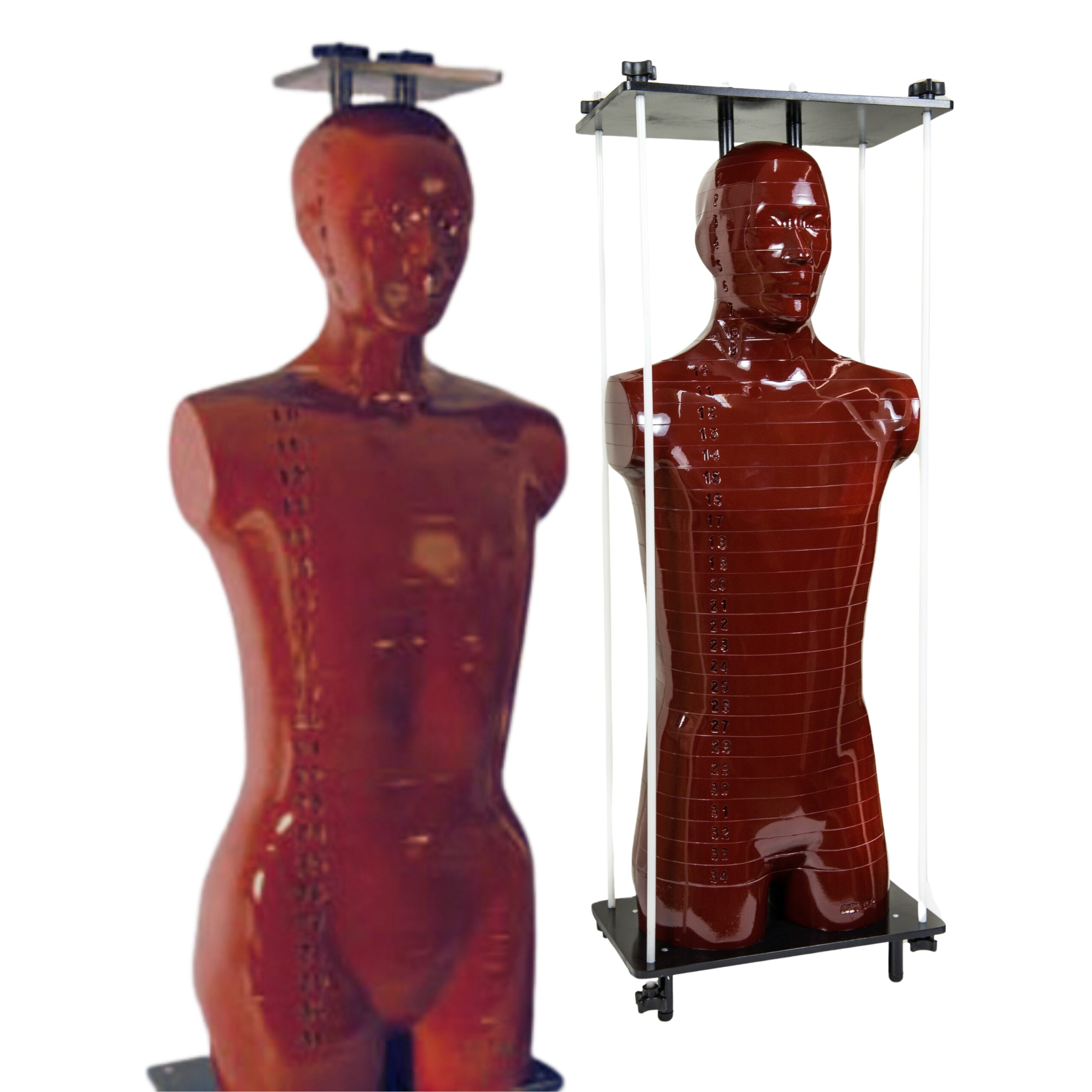
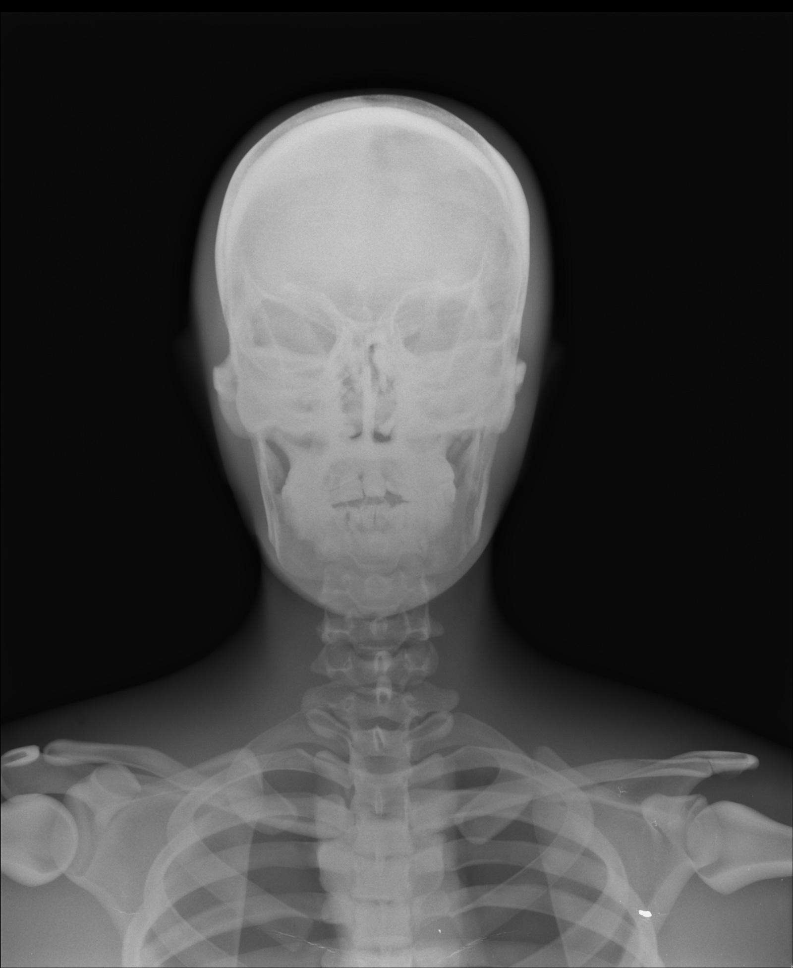
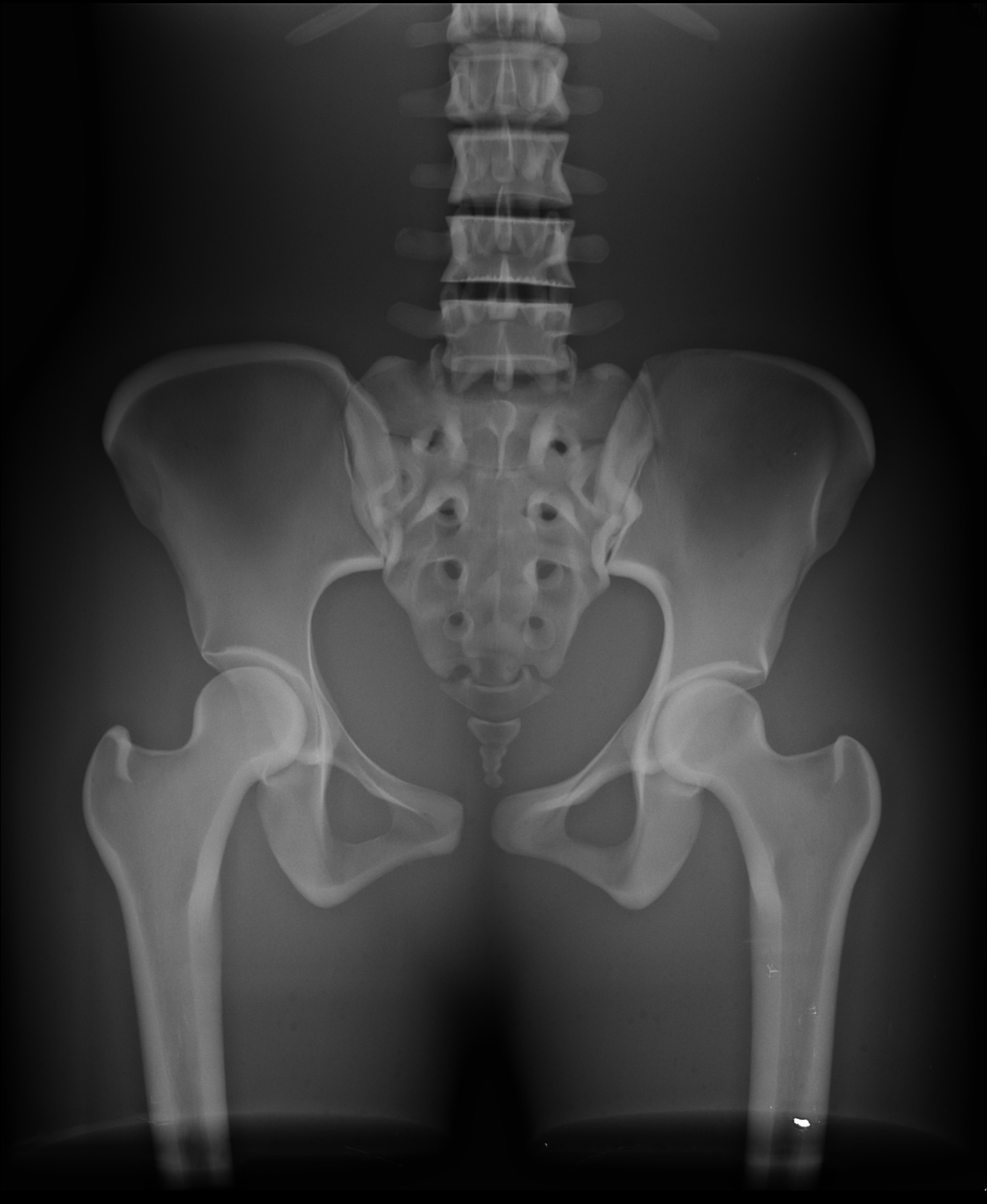
Building on decades of clinical trust, RSD’s ART Phantom with Cadaver Bones incorporates authentic human skeletal material to replicate real tissue density, bone structure, and attenuation characteristics with a fidelity that synthetic substitutes cannot match. The result: QA data your team can trust, from simulation through final delivery.
Engineered as a refined evolution of the Alderson RANDO Phantom, our ART Phantom with Cadaver Bones is designed within precise technological constraints and manufactured in full conformance with ICRU-44 standards. It provides integrated testing of the entire treatment planning and delivery chain – from imaging and contouring to dose calculation and plan execution – in a single, streamlined workflow.
True-to-anatomy accuracy. Built-in end-to-end verification. Designed for the demands of modern radiation therapy.
Advantages of Cadaver Bones
Synthetic bone substitutes may introduce known discrepancies in Hounsfield unit values, beam attenuation, and scatter characteristics. For institutions performing high-precision treatments, these gaps matter.
Real cadaver bone provides the structural heterogeneity, density gradients, and material properties found in your actual patients. The result: phantom-based QA that more faithfully mirrors clinical conditions, giving your team greater confidence in treatment plan verification and imaging system performance.
Anatomy
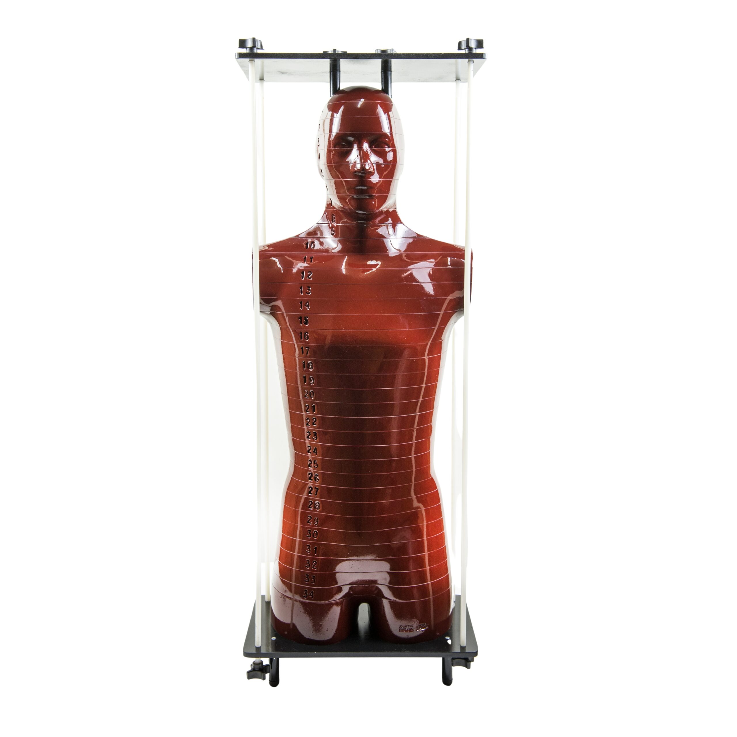
The male ART Phantom represents a 175 cm (5 ft. 9 in.), 73.5 kg (162 lb.) adult male; the female ART Phantom represents a 155 cm (5 ft. 1 in.), 50 kg (110 lb.) adult female. Both phantoms now incorporate authentic cadaver bones, replacing synthetic bone-equivalent inserts with real skeletal material for accurate Hounsfield unit representation and true heterogeneous attenuation.
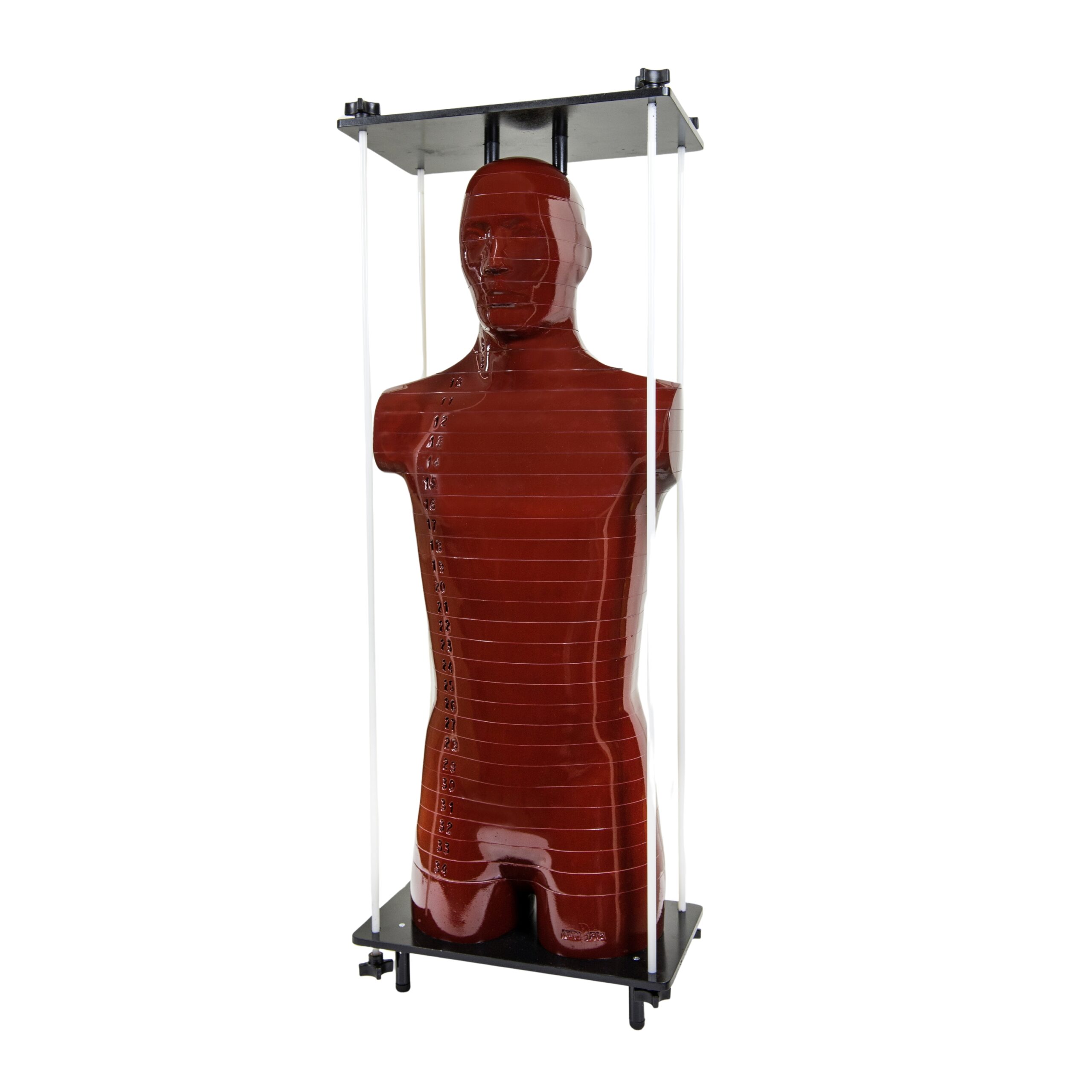
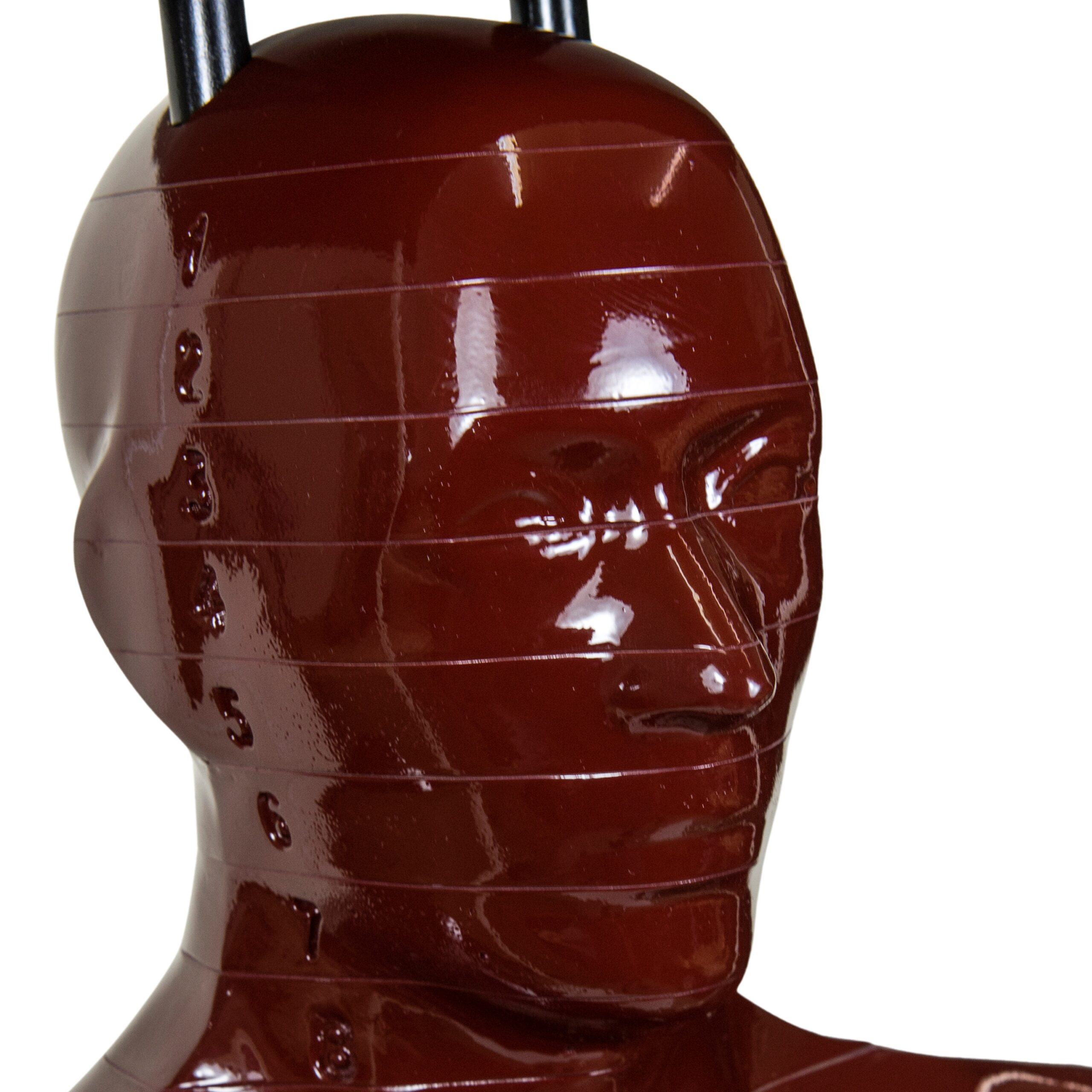
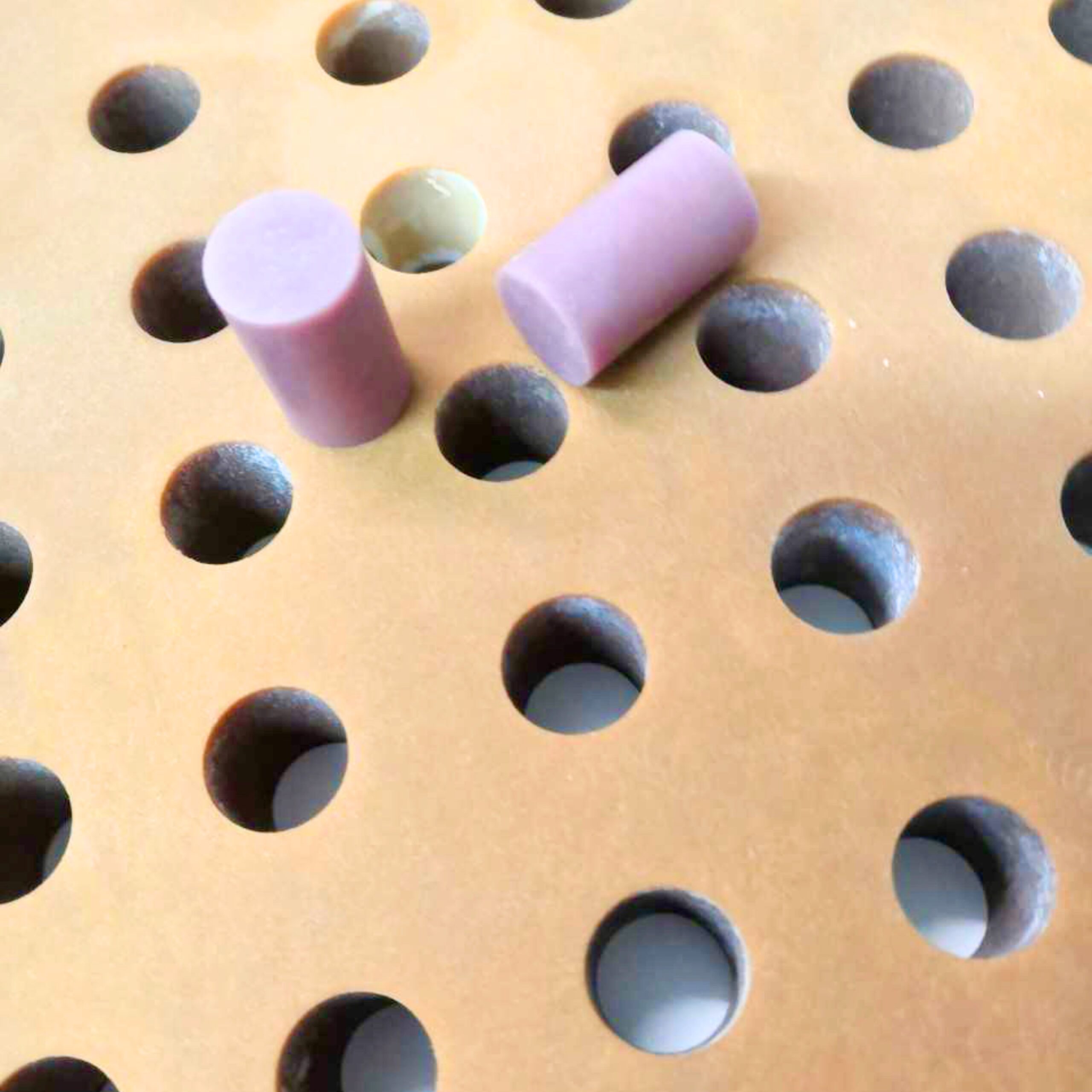
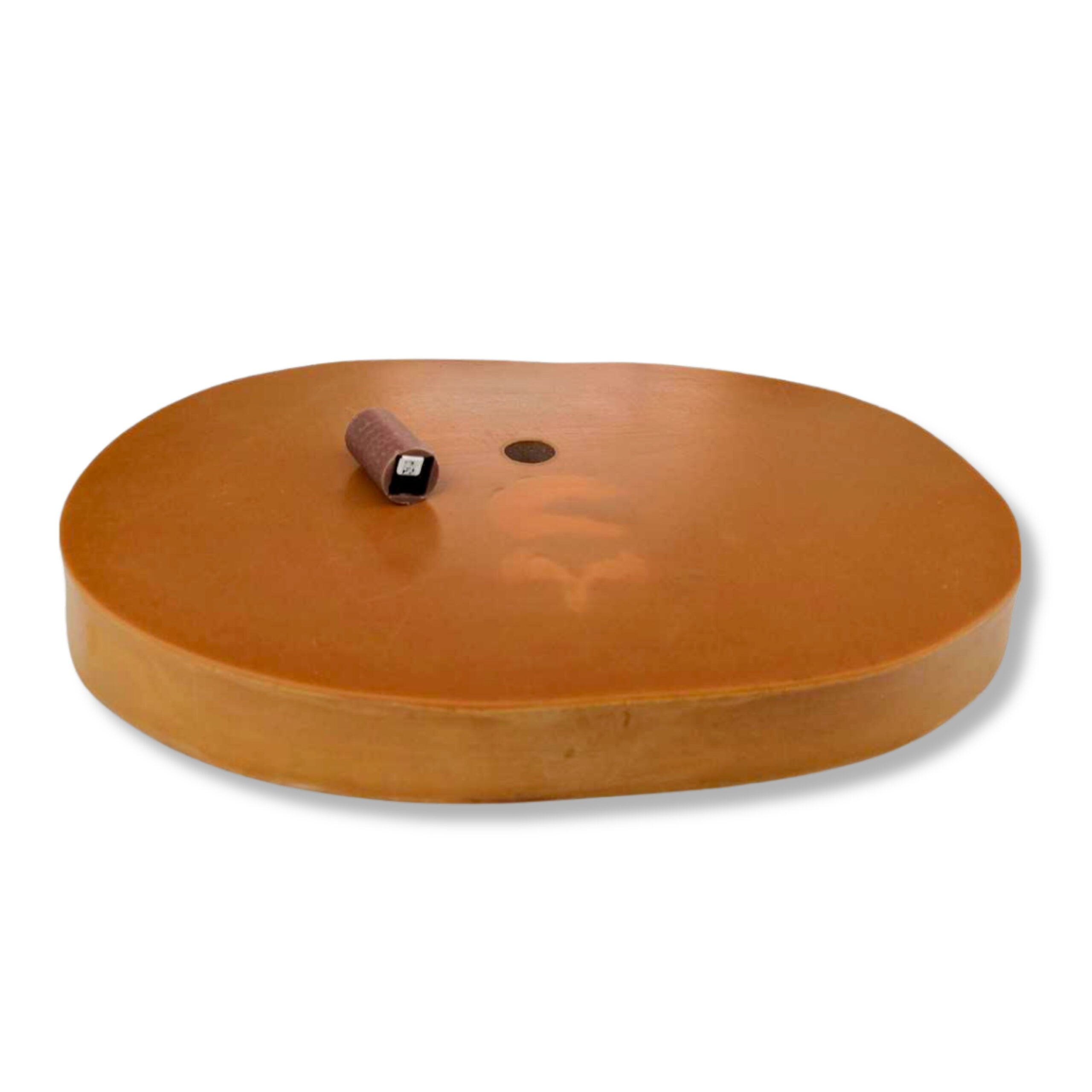
Each phantom is sectioned transversely into 2.5 cm thick slices. Slices are fitted with interchangeable pins fabricated from bone-equivalent, so!-tissue-equivalent, or lung-tissue-equivalent materials. All pins are replaceable with TLD holder pins to accommodate thermoluminescent dosimetry; holder pins are available as a separate order item.
Slice surfaces are finished with a so!-tissue-equivalent coating, producing glass-smooth interfaces for precise geometric registration between sections. Coatings are selectively removed over the air spaces of the oronasal pharynx, trachea, and stem bronchi to preserve anatomically accurate airway geometry.
Dosimetry holes are drilled in 3 cm × 3 cm or 1.5 cm × 1.5 cm grids at diameters of 5 mm and 7 mm, providing a high-resolution measurement matrix for detailed mapping of three-dimensional dose distributions throughout the phantom volume.
Materials
Soft Tissue: The human body exhibits continuous, small variations in soft-tissue density and attenuation throughout. The ART Phantom’s so!-tissue-equivalent material is closely controlled to replicate the mean density of human so! tissues, in conformance with ICRU Report 44 specifications.
Skeleton: This configuration incorporates authentic human cadaver bones, selected and prepared to match the anthropometric dimensions of the phantom’s soft-tissue molds. Cadaver bone provides accurate cortical bone density, intact trabecular (spongiosa) microarchitecture, and preserved marrow spaces – material properties that cannot be fully reproduced by synthetic bone-equivalent substitutes.
The use of real skeletal material eliminates discrepancies in Hounsfield unit accuracy, heterogeneous attenuation, and cortical-to-trabecular density gradients that are inherent to polymer moldings, making this configuration the appropriate choice for dosimetric applications requiring the highest anatomical fidelity.
Lungs: Lung volumes are molded from syntactic foam with a specific gravity of 0.30 g/cc, providing a reproducible, low-density tissue-equivalent material consistent with average in vivo lung density.
TLD Dosimeters and Fittings
All phantoms are shipped with dosimetry holes filled with blank pins. Standard pins are 2.50 cm in length unless otherwise specified.
TLD chip holder pins feature a recess measuring 3.2 × 3.2 × 0.9 mm at one end. TLD rod holder pins are fitted with a 1 mm-diameter cross-drilled hole at the pin center. Pins are also available, configured to accommodate OSLD dosimeters of various types.
Tissue-equivalent plugs are available machined to fit the following detector formats: TLD chips, TLD rods, TLD bars, TLD cubes, MOSFET detectors, LANDAUER® OSL MicroSTAR® holders, LANDAUER® nanoDot® holders. Holder pins for all detector formats are available separately.
LANDAUER®, MicroSTAR®, and nanoDot® are registered trademarks of LANDAUER®, a division of Fluke® Corporation.
Assembly
ART Phantom slices are secured between aluminum end plates using nylon tie rods with tightening knobs that clamp slices firmly in proper anatomical alignment. Both internal and external assembly configurations are included with the phantom.
The external assembly is designed to facilitate film dosimetry, providing unobstructed slice interfaces for planar dose measurement. The internal assembly is intended for use with TLD or ion chamber dosimetry, supporting point-dose and volumetric measurements within the phantom volume.
Breast Attachments
Breast attachments are available in various sizes, contoured to blend realistically with the thorax of both male and female ART Phantoms, and secured with nylon screws. The male chest with breast attachments fitted serves as a large female configuration.
Breasts may be sectioned in frontal planes and are available drilled or undrilled for film dosimetry. Sliced breasts accept the full range of tissue-equivalent pins described in the TLD Dosimeters and Fittings section.
To order breast attachments, please specify the following:
Product Gender:
ART-250 | Male Breasts
ART-350 | Female Breasts
Volume:
| Male Breast Attachments | Volume |
| 250 ml | |
| 500 ml | |
| 750 ml | |
| 1,000 ml | |
| 1,250 ml | |
| Female Breast Attachments | Volume |
| 200 ml | |
| 300 ml | |
| 400 ml | |
| 500 ml | |
| 650 ml |
Sliced or Unsliced:
-S | Sliced
-U | Unsliced
Hole Grid:
-1.5 | 1.5cm x 1.5cm
-3.0 | 3.0cm x 3.0cm
-0 | None
Hole Size:
-5 | 5mm Diameter
-7 | 7mm Diameter
Side:
-L | Left
-R | Right
-P | Pair
Example:
ART-250 – 500 – S – 3.0 – 7 – P
Male Breast – 500ml – Sliced – 3.0cm x 3.0cm Grid – 7mm Diameter – Pair
Model Numbers
| UNDRILLED | 3 cm x 3 cm GRID HOLE SPACING | 1.5 cm x 1.5 cm GRID HOLE SPACING | |
| Male ART Phantom with Cadaver Bones (Sections 0-35) | ART-200XCB | ART-200CB | ART-200ACB |
| Female ART Phantom with Cadaver Bones (Sections 0-32) | ART-300XCB | ART-300CB | ART-300ACB |
Applications
- Organ specific dosimetry for all dosimeters (TLD, OSL nanodots, MOSFET, film, ion chambers, and diodes)
- Standard 3 cm x 3 cm or 1.5 cm x 1.5 cm hole grids for dosimeters
- IMRT organ dose distributions
Modalities
- External beams in the 0.04 to 40 MeV
- Intensity-Modulated Radiation Therapy (IMRT)
- Stereotactic Body Radiation Therapy (SBRT)
- Gamma Knife
- CyberKnife
- CT
- Cone Beam CT
Clinical Images

Publication References
Flatten V, Friedrich A, Engenhart-Cabillic R, Zink K. A Phantom Based Evaluation of the Dose Prediction and Effects in Treatment Plans, When Calculating on a Direct Density CT Reconstruction. J Appl Clin Med Phys. 2020 Mar;21(3):52-61. PMID: 32176455; PMCID: PMC7075385. DOI: 10.1002/acm2.12824
Hauri P, Schneider U. Whole-Body Dose Equivalent Including Neutrons is Similar for 6 MV and 15 MV IMRT, VMAT, and 3D Conformal Radiotherapy. J Appl Clin Med Phys. 2019 Mar;20(3):56-70. Epub 2019 Feb 21. PMID: 30791198; PMCID: PMC6414138. DOI: 10.1002/acm2.12543
Sawyer L J, Whittle S A, Matthews E S, Starritt H C, JUPP T P. Estimation of Organ and Effective Doses Resulting From Cone Beam CT Imaging for Radiotherapy Treatment Planning. British Journal of Radiology, Vol. 82, No. 979. 2014 Mar. DOI: 10.1259/bjr/62467578