


NEW! Head Phantom with Cadaver Bones
PROFESSIONAL TRAINING AID WITH CADAVER BONE CONSTRUCTION FOR RADIOLOGICAL TECHNOLOGISTS
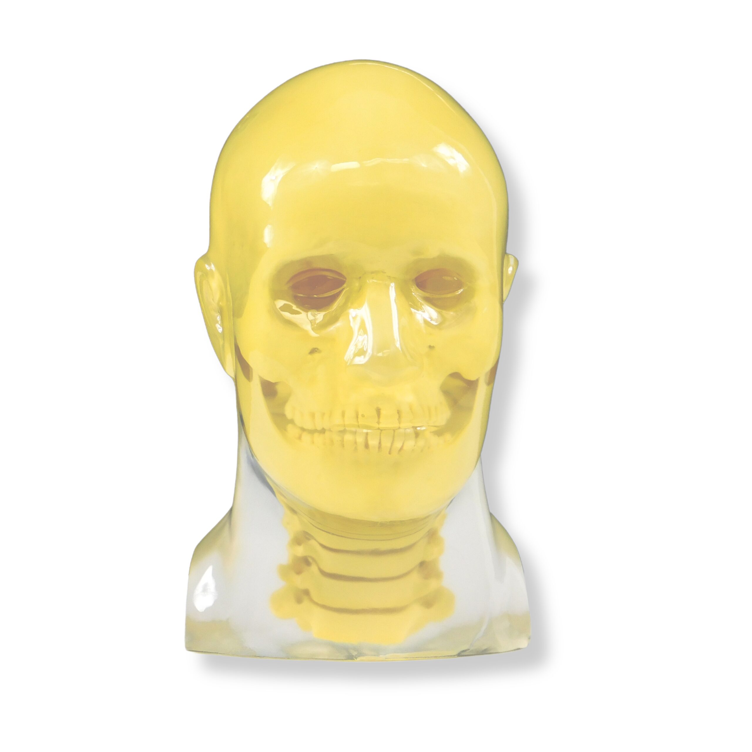
Train with confidence using RSD’s Head Phantom, purpose-built to deliver realistic, clinically relevant practice for radiological technologists at every level. Combining the acclaimed anatomy and radiological fidelity of RSD’s Take-Apart Pixy, this phantom represents an average adult male – 5’9″ (175 cm), 162 lbs (74 kg) – giving learners a true-to-life training experience. Built for flexibility. Designed for real-world training.
Choose the configuration that fits your program’s needs:
- Opaque or transparent construction
- With or without cervical spine
- Multiple skin tones for inclusive, representative training
The Head Phantom is rugged, shatter-proof, and easy to transport, so it holds up in busy lab environments and travels wherever your training takes you. A step beyond geometric phantoms.
RSD Body Sections aren’t a replacement for simple geometric phantoms used to evaluate individual imaging system characteristics. They go further, providing comprehensive, realistic assessment of both imaging systems and imaging techniques under true-to-life conditions. Because great radiological technologists train on the real thing.
Advantages of Cadaver Bones
Synthetic bone substitutes may introduce known discrepancies in Hounsfield unit values, beam attenuation, and scatter characteristics. For institutions performing high-precision treatments, these gaps matter.
Real cadaver bone provides the structural heterogeneity, density gradients, and material properties found in your actual patients. The result: phantom-based QA that more faithfully mirrors clinical conditions, giving your team greater confidence in treatment plan verification and imaging system performance.
Model Numbers
| RS-108CB | Head Phantom with Cadaver Bones and Cervical Spine | Opaque |
| RS-108TCB | Head Phantom with Cadaver Bones and Cervical Spine | Transparent |
| RS-109CB | Head Phantom with Cadaver Bones | Opaque |
| RS-109TCB | Head Phantom with Cadaver Bones | Transparent |
Please contact RSD for custom pathologies and traumas.
Applications
- Teaching & training
- Image quality
- Panographic Imaging
- Dosimetry verification
- Protocol verification
Modalities
- CT
- X-Ray
- Fluoroscopy
- Dental X-Ray
- CBCT
Anatomy
- Authentic human cortical and trabecular bone providing true-to-life radiological density and anatomical accuracy
- Brain material composed of RSD ART soft tissue material (TS-1001-T)
- Spinal cord material made of ART soft tissue material with density of 1.1 g/cc
- Oral, trachea, and sinus cavities filled with Styrofoam
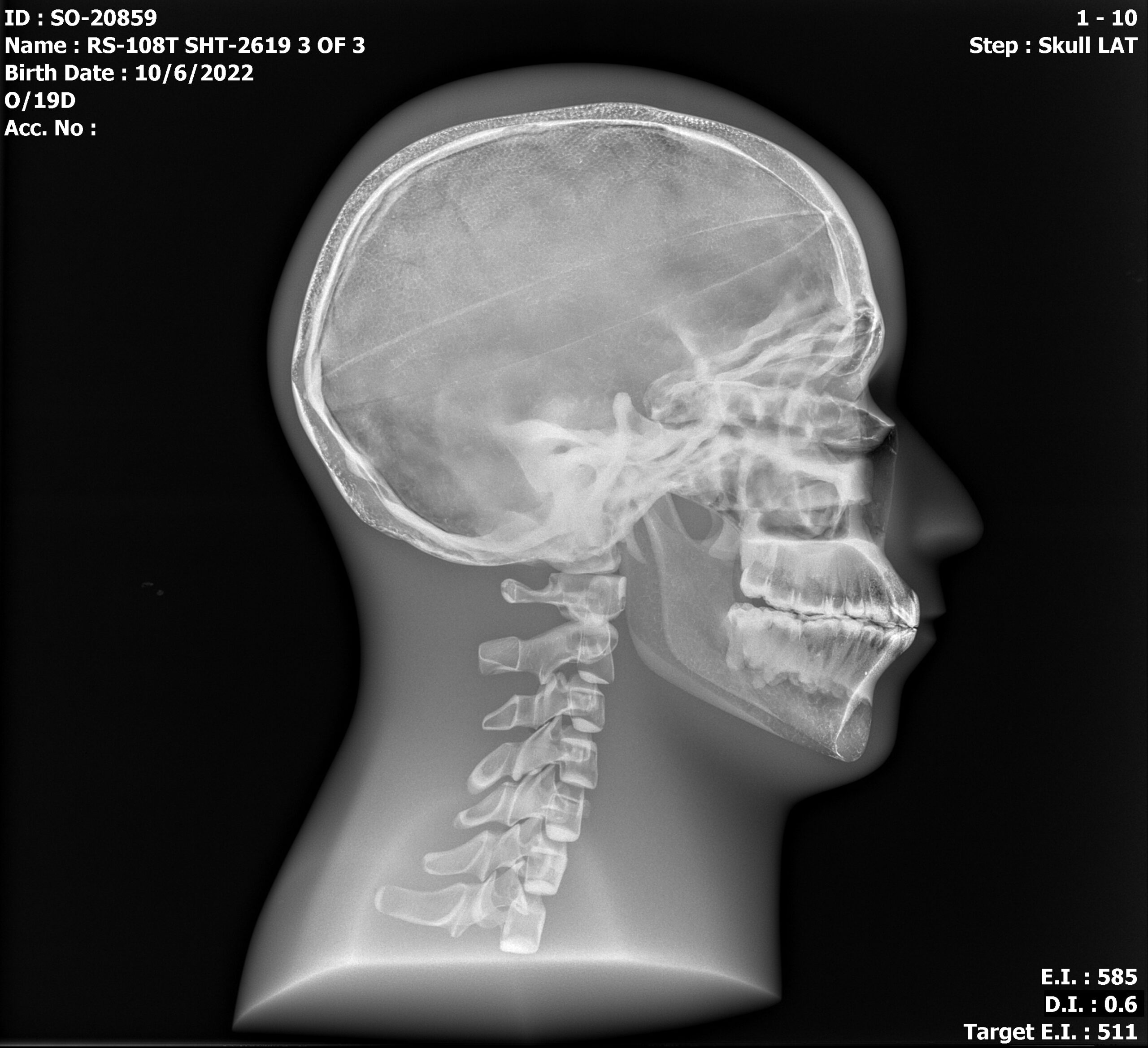
Clinical Images
Publication References
Compagnone G, Pagan L, Bergamini C. Comparison of Six Phantoms for Entrance Skin Dose Evaluation in 11 Standard X-Ray Examinations. J Appl Clin Med Phys. 2005;6(1):101-113. DOI: 10.1120/jacmp.v6i1.2020
Akyalcin S, English JD, Abramovitch KM, Rong XJ. Measurement of Skin Dose From ºCone-Beam Computed Tomography Imaging. Head Face Med. 2013;9:28. Published 2013 Oct 9. DOI: 10.1186/1746-160X-9-28