
Domestic Violence Take-Apart Pixy
ANTHROPOMORPHIC TRAINING & TEACHING PHANTOM WITH 16 PATHOLOGIES AND TRAUMAS
- Phantom disassembles into 9 parts
- Ideal substitute for teaching and training radiological technologists
- Small size and low weight simplify positioning
- Unlimited repetition of most views for which patients cannot be used
- Provides valid feedback to evaluate trainee performance
- Designed to image any clinical view (AP, oblique, lateral, frog legs, etc.)
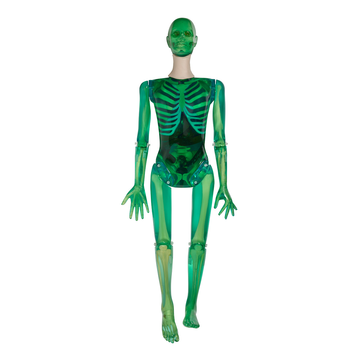
Designed with the expert assistance of Judy McNitt-Mell, RSD’s Domestic Violence Take-Apart Pixy is an anatomically and radiologically correct female designed specifically for training radiologic technologists. At 5’ 1” (156 cm) weighing 105 lbs (48 kg), Domestic Violence Take-Apart Pixy is a repeatable, convenient substitute for patients and virtually indestructible.
Domestic Violence Take-Apart Pixy is tinted green with visible skeleton and organs, including the stomach, gall bladder, urinary bladder, kidneys, rectum, and sigmoid flexure. The organs are air-filled but accept water or contrast media that can be easily flushed after use. Custom fractures and pathologies are available at an additional cost.
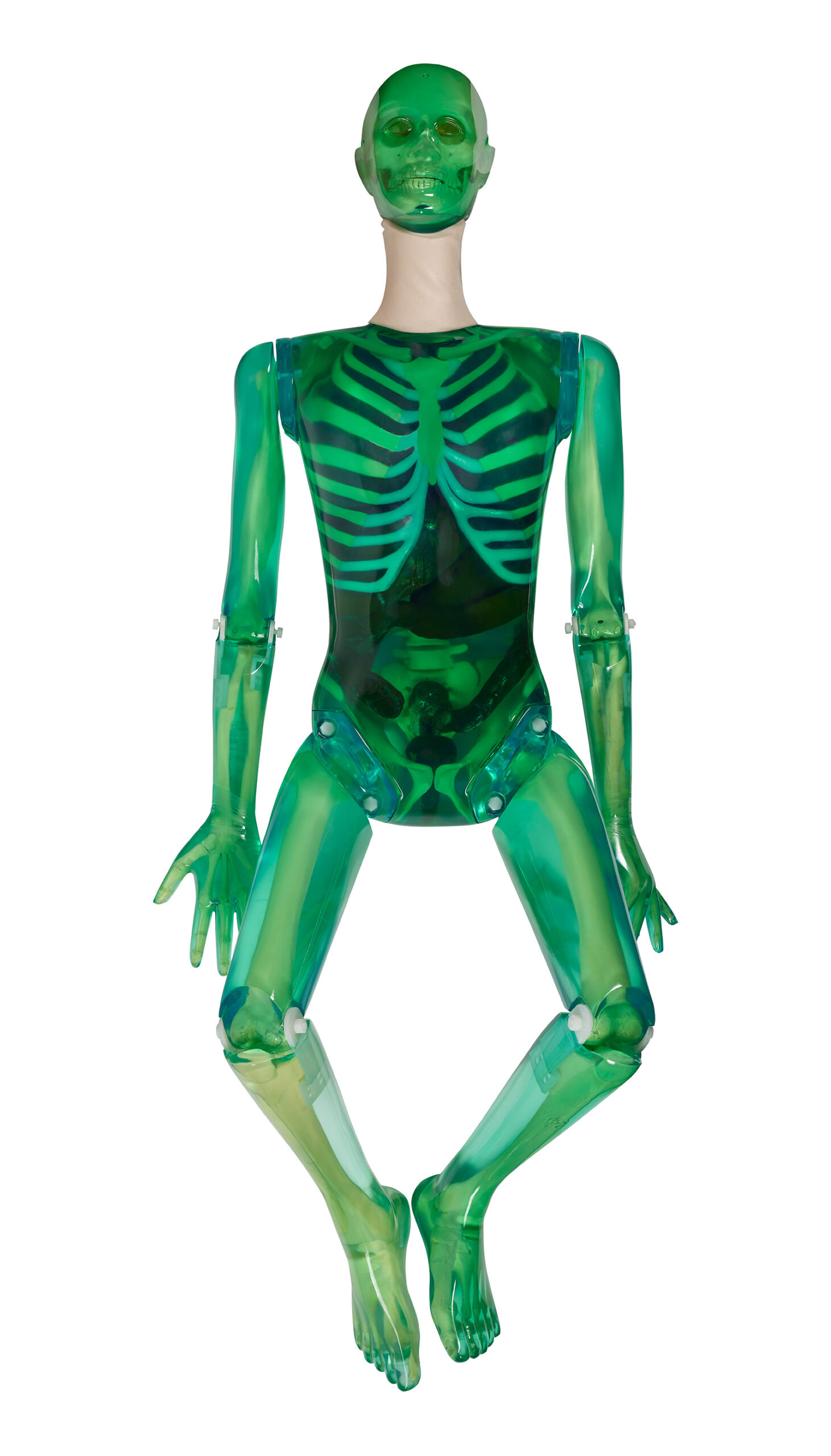
Built with soft-tissue mold and skeleton molds that are matched for anatomic fidelity, Domestic Violence Take-Apart Pixy permits unlimited exposures, demonstrates the effects of changing technical factors, and allows for the evaluation of student performance. Students have no difficulty in maneuvering Domestic Violence Take-Apart Pixy into most desired positions as the phantom is built to tolerate trainee errors.
Domestic Violence Take-Apart Pixy is used to demonstrate anatomy and evaluate positioning and imaging techniques, including kVp, mAs, contrast, optical density, digital processing, OFD, and TFD. Made of tissue-equivalent materials and life-like articulations, Domestic Violence Take-Apart Pixy is more realistic than a cadaveric skeleton with radiographs that are optically equivalent in density and contrast to human patients.
C1, C2, and C7 were converted to mechanical nylon joints because educators in the field prefer full positioning capabilities for the head. This design permits the remaining neck vertebrae to be fixed in a normal position while assuring a full range of head motion.
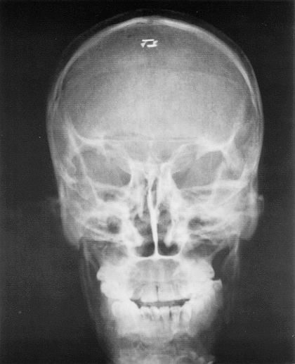
The skull of Domestic Violence Take-Apart Pixy has frontal and sphenoidal sinuses, ethmoidal and mastoid air cells, and the auditory ossicles. Bone sutures are radiographically visible.
Domestic Violence Take-Apart Pixy lungs are molded of tissue-equivalent foam with the mass density of inflated human lungs (0.30 g/cc). They are connected to the oro-nasal cavity by the stem bronchi and trachea. The oro-nasal pharynx is filled with a nearly air-equivalent foam.
Model Number
| RS-106T | Transparent | Domestic Violence Take-Apart Pixy with NO Fill Ports. Includes storage case. |
Applications
- ER evaluation of technologist performance in identifying common trauma of domestic violence
- Teaching & training of patient positioning
- Image quality
- Diagnostic radiology
- Dosimetry verification
- Protocol verification
Modalities
- CT
- X-Ray
- Fluoroscopy
Anatomy
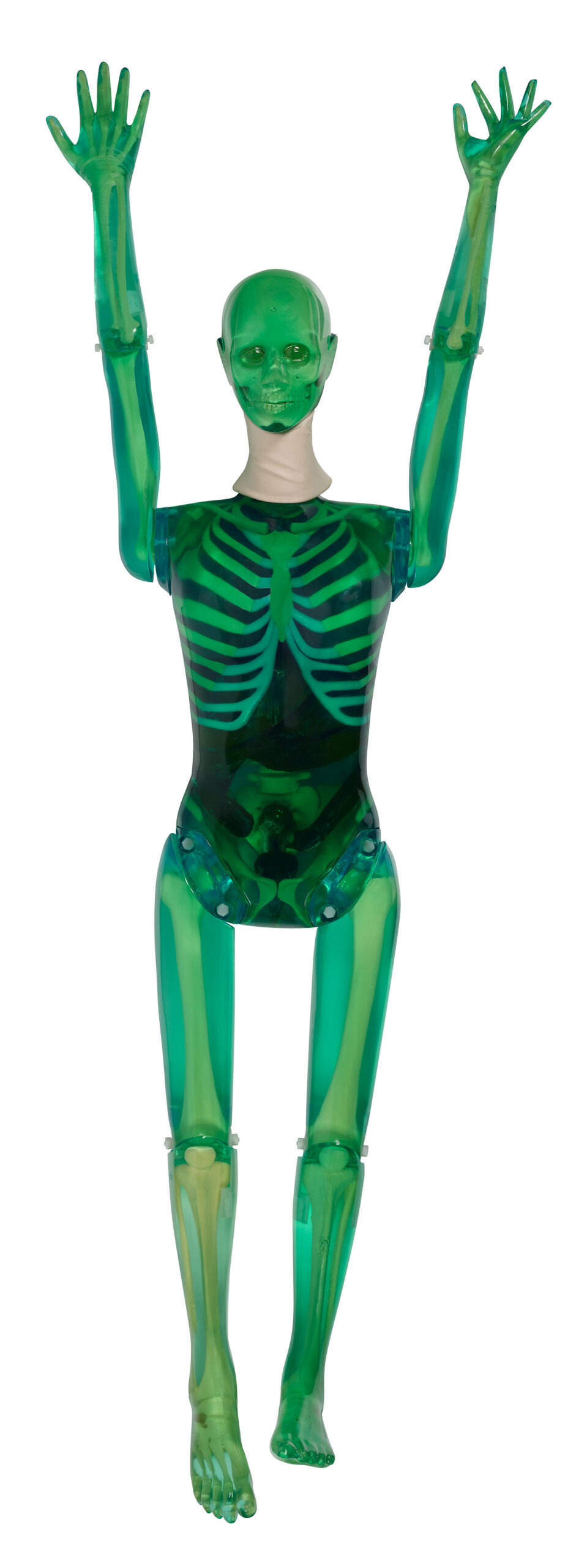
- Shoulders have ball and socket joints
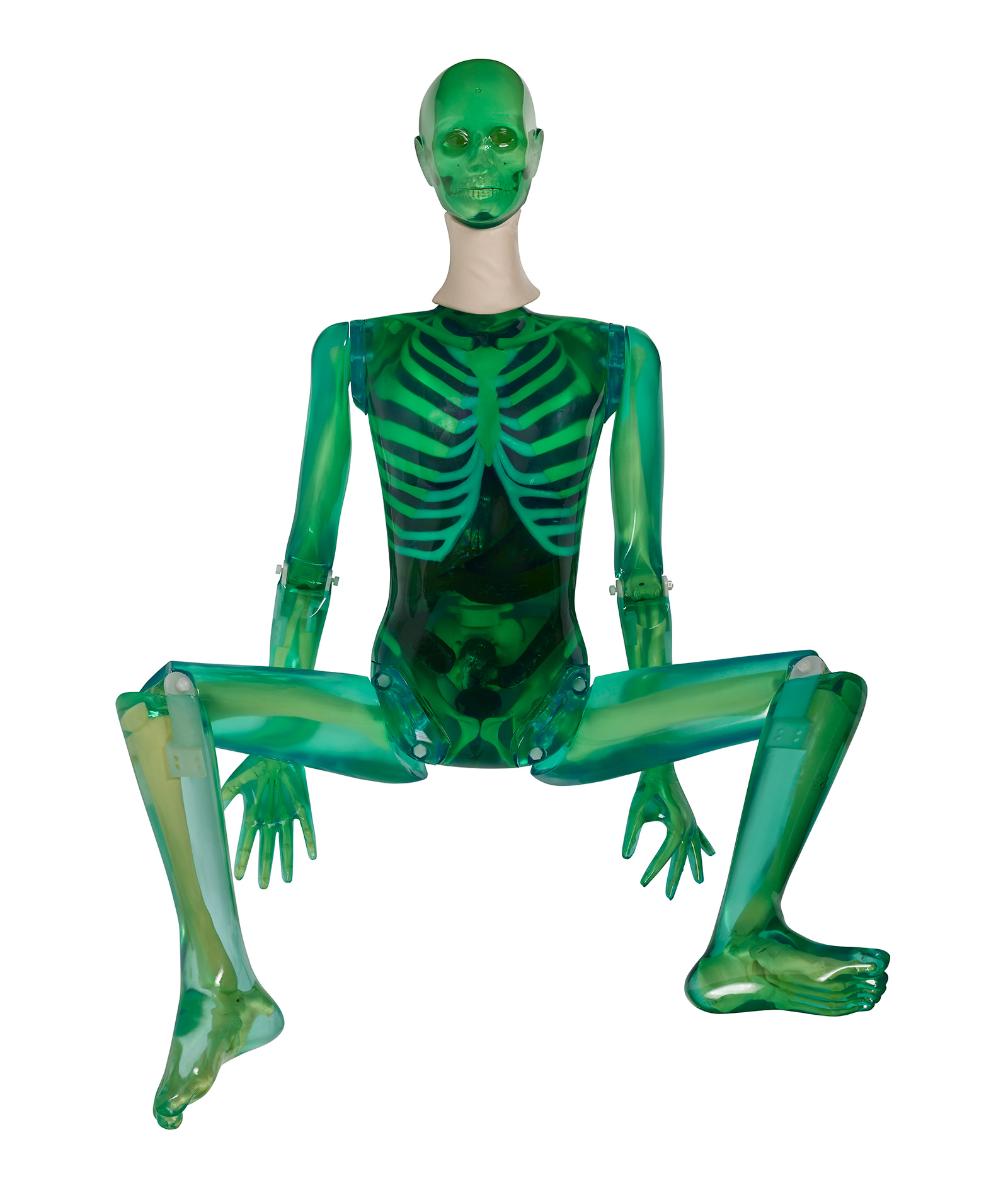
- Elbows and knees flex 90° to 100°
- A “frog position” is possible at the hips
- Right hand is molded with fingers positioned for an AP view
- Left hand is in an oblique-lateral position
- Feet are in natural position
Clinical Images
Pathologies & Traumas
| Head | Displacement of mandibular condyle fracture Separate fracture of left frontal zygomatic bone Step deformity of left infraorbital rim Mandible fracture with missing bone Fracture of nasal bone |
| Neck | C4+C5 compression fracture |
| Body | Glenoid Fracture left scapular Fracture of lateral right ribs 6+7 Fracture of left ribs 8+9 mediolateral Right 12 rib fracture |
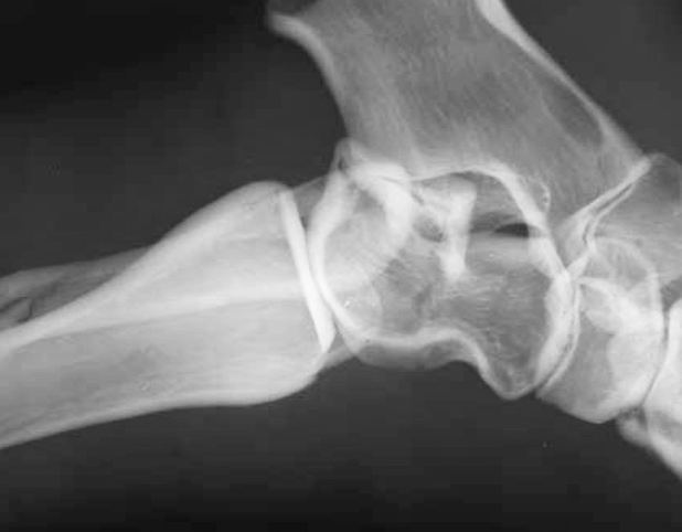
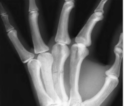
| Arm | Displaced fracture of left radius and ulna, midshaft Left hand 3rd and 4th metacarpals shattered Proximal tibia fracture with multiple bone fragment Right minimally displaced distal fibula fracture |
| Pelvis | Displaced pubic ramus fracture right and left |
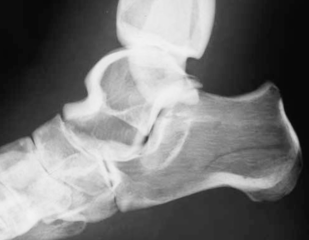
| Foot | Right displaced angle fracture in calcaneus bone |
RSD In Action
Keele University’s School of Health and Rehabilitation opened a state-of-the-art radiography suite using RSD’s PIXY Phantom to highlight the practice of hands-on training with visual reinforcement.