

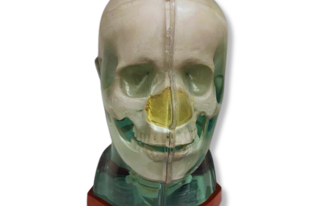
You likely already know that a phantom is a stand-in for a human head during scans. But the real question isn’t just what is a head phantom for medical imaging, but what makes one truly effective for today’s advanced applications? The answer goes far beyond simple geometric shapes for basic tests. Modern phantoms offer incredible anatomical accuracy, with some incorporating real human bone to provide an unmatched level of realism for complex studies. They are also designed for multi-modality use, allowing you to correlate data from CT, MRI, and PET scans of the same object. This article moves past the basics to explore the critical features that define a high-quality phantom and how to choose the right one for demanding research, AI validation, and therapy planning.
Key Takeaways
- Establish a reliable quality control baseline: Head phantoms provide a consistent benchmark for calibrating your equipment, helping you verify that every scan is safe and accurate before any patient interaction.
- Match the phantom type to your specific goal: Select a static phantom for basic calibration, a dynamic one to simulate patient movement, or a multi-modality model to compare results across different imaging systems like CT and MRI.
- Prioritize anatomical accuracy and versatility: Choose a phantom with realistic structures and tissue-equivalent materials for clinically relevant results, and consider multi-modality options to streamline testing across different platforms.
What Is a Medical Imaging Head Phantom?
So, what exactly is a medical imaging head phantom? Think of it as a highly specialized stand-in for a human head. It’s a physical or computational model designed to test, calibrate, and improve the performance of medical imaging equipment. These tools are essential for ensuring that machines like CT scanners, MRI systems, and X-ray units are working correctly before they are ever used on a patient. Phantoms allow physicists, technicians, and researchers to perform repeatable tests in a controlled environment, something that simply isn’t possible with a living person.
These models range from simple geometric shapes used for basic quality checks to incredibly detailed anthropomorphic phantoms that mimic the complex anatomy of the human head. The primary goal is to simulate how different tissues and structures within the head interact with various imaging technologies. By using a phantom with known properties, you can precisely measure image quality, assess radiation dosage, and validate new imaging techniques. This makes them an indispensable part of quality assurance programs, medical training, and cutting-edge research in fields from neurology to radiation oncology. Essentially, they provide a reliable benchmark, giving you the confidence that your imaging systems are delivering accurate and safe results for every patient, every time.
How phantoms replicate human tissue
A phantom’s main job is to act like human tissue when it’s being scanned. These models are carefully engineered to simulate the physical properties of different tissues, such as bone, brain matter, and soft tissue, for all kinds of medical imaging modalities. This includes everything from conventional radiography and CT to MRI and ultrasound. The materials used are chosen for their ability to absorb or scatter radiation, or react to magnetic fields, in a way that is equivalent to real human tissue.
While some phantoms use biological materials, they can be difficult to preserve and often lack consistency over time. This is why manufactured phantoms are so valuable. They provide a stable, reliable, and reproducible standard. This consistency ensures that the tests you run today will be comparable to the tests you run months or even years from now, giving you confidence in your equipment’s performance and the accuracy of your data.
The importance of anatomical accuracy
Beyond just mimicking tissue properties, a phantom must also be anatomically accurate. Anthropomorphic phantoms are designed to replicate the specific shapes, sizes, and spatial relationships of structures within the human head. This level of detail is critical for conducting clinically meaningful imaging studies. For example, a phantom with a realistic skull, brain, and sinus cavities allows you to test how well an imaging system can distinguish between different structures.
This accuracy is essential for everything from planning complex surgeries to developing new treatment protocols. Phantoms with known geometries and material compositions allow you to precisely characterize an imaging system’s ability to produce safe and accurate images. When a phantom includes fine details, like those found in our Multi-Modality Phantoms, it provides a much more rigorous and realistic test for your equipment, helping you refine techniques and train staff for real-world clinical scenarios.
Physical vs. Computational Head Phantoms
When we talk about head phantoms, it’s helpful to know they come in two main forms: physical and computational. Each type serves a distinct purpose in medical imaging and therapy, and sometimes they even work together. Think of it as having both a physical model and a digital blueprint. A physical phantom gives you a tangible object to scan and test, while a computational phantom provides a flexible, data-driven model for simulations. Understanding the difference helps you select the right tool for calibrating equipment, training staff, or conducting research. As technology advances, the line between these two is also starting to blur, creating exciting new possibilities for medical physics.
Understanding physical phantoms
Physical phantoms are tangible, three-dimensional objects designed to mimic the size, shape, and tissue properties of a human head. You can think of them as sophisticated stand-ins used to test and calibrate imaging machines in a controlled, repeatable way. These models are built with materials that have known characteristics, allowing you to precisely check how well an imaging system can produce accurate images. By using a physical phantom, you can test equipment performance without exposing a real person to radiation. This makes them essential tools for routine quality assurance and for developing new imaging techniques safely and effectively.
Exploring computational (virtual) phantoms
Computational phantoms, also known as virtual phantoms, are detailed computer models of the human body. Instead of a physical object you can hold, these are data files that can be loaded into software for simulations. They are incredibly useful for planning treatments like radiation therapy and for studying how different imaging techniques work. For example, a computational phantom can include a digital representation of a tumor, allowing researchers to test an imaging system’s ability to detect it. Because they are digital, these models can be easily modified to represent different anatomies or conditions, offering great flexibility for research and planning.
Hybrid phantoms: Bridging the physical and digital
The latest innovations in phantom technology are starting to combine physical and computational models to create hybrid solutions. These “digital twin” phantoms link a physical model to a real-time computer simulation. Imagine a physical head phantom that can simulate changes like tumor growth or organ movement through its connected digital twin. This approach adds a dynamic layer to testing, making it possible to evaluate how well AI imaging systems can predict and track changes over time. By merging the tangible feedback of a physical object with the flexibility of a digital simulation, these advanced multi-modality phantoms are expanding what’s possible in medical research and validation.
Common Types of Physical Head Phantoms
Physical head phantoms come in several forms, each designed for a specific purpose. The main categories are static, dynamic, and multi-modality. Choosing the right one depends entirely on what you need to accomplish, whether that’s calibrating a machine for a stationary target, evaluating its performance with physiological motion, or ensuring consistency across different imaging systems. Understanding the differences will help you select the most effective tool for your quality assurance, research, or training needs.
Static head phantoms
Static head phantoms are designed to be completely still, representing a fixed anatomical state. These are your go-to tools for fundamental tasks like equipment calibration and testing spatial resolution. Because they don’t move, they provide a stable and repeatable target for training staff on how to operate scanners or for testing how well a system can visualize stationary objects. For example, a static phantom is perfect for practicing the detection of foreign bodies like shrapnel or small bone fragments. They establish a reliable baseline for image quality, making them an essential part of any medical imaging department’s quality assurance protocol.
Dynamic head phantoms
Unlike their static counterparts, dynamic head phantoms are built to simulate physiological movement. Think of the subtle motion of breathing, the pulsation of blood flow, or even small patient movements. These phantoms incorporate mechanisms that replicate these real-world conditions, allowing you to assess how well an imaging system performs when things aren’t perfectly still. This is especially important for advanced imaging techniques and for planning treatments in radiation therapy, where targeting accuracy is critical despite patient motion. Using a dynamic phantom helps ensure your imaging modality can capture clear, accurate images under realistic clinical scenarios.
Multi-modality head phantoms
Multi-modality head phantoms are the versatile workhorses of the phantom world. They are constructed from materials that are visible across different imaging systems, such as CT, MRI, and PET. This allows you to image the same phantom on multiple machines to compare and correlate the results, which is vital for image fusion studies. Beyond quality assurance, these phantoms are increasingly used to train and validate AI algorithms for tasks like image segmentation and automated diagnosis. RSD offers a variety of multi-modality phantoms that provide the consistency needed for both routine QA and advanced research applications.
How Different Imaging Modalities Use Head Phantoms
Head phantoms are essential across all types of medical imaging, but their specific role changes depending on the technology. Each imaging modality, from CT to MRI, interacts with human tissue in a unique way. Because of this, phantoms are designed to test the specific principles of each system. Whether it’s measuring radiation dose, checking magnetic field uniformity, or tracking a radiotracer, a head phantom provides a stable and reliable subject. This allows technologists and physicists to fine-tune their equipment and procedures, ensuring every scan is as safe and accurate as possible. By simulating the complexities of the human head, these tools help teams verify that their systems are performing correctly before they are ever used on a patient.
CT and X-ray
In computed tomography (CT) and X-ray imaging, phantoms are fundamental to quality control. These modalities use ionizing radiation to create images, so it’s critical to ensure the equipment is calibrated perfectly. A head phantom allows you to verify that the system is operating correctly, helping you optimize imaging protocols to achieve the clearest possible images with the lowest radiation dose. By scanning a phantom with known densities and structures, you can check for artifacts, assess image resolution, and confirm dose accuracy. This routine testing is a cornerstone of any quality assurance program, providing confidence that your equipment is both effective and safe for patient use.
MRI
Magnetic Resonance Imaging (MRI) uses powerful magnetic fields and radio waves instead of ionizing radiation. For this modality, head phantoms provide a controlled and reproducible framework for evaluating the technology. They are used to test everything from the uniformity of the magnetic field to the performance of new imaging sequences. Because phantoms simulate the magnetic properties of different human tissues, they offer a reliable way to assess image quality, geometric accuracy, and signal-to-noise ratio without the variables that come with a human subject. This makes them invaluable for routine quality checks, system acceptance testing, and research into new MRI techniques. Many facilities use multi-modality phantoms that work for both MRI and CT.
Nuclear medicine and PET
For nuclear medicine and Positron Emission Tomography (PET), which use radiotracers to visualize metabolic processes, head phantoms play a dual role. First, they are used to calibrate scanners and verify that the equipment can accurately detect and quantify radioactive signals. Second, they are becoming increasingly important for training and validating machine learning algorithms. Phantoms provide consistent, customizable ground-truth datasets that are perfect for teaching AI to recognize disease patterns, segment anatomical structures, and improve diagnostic accuracy. This application is especially critical in nuclear medicine, where quantitative accuracy directly impacts treatment planning and monitoring.
Ultrasound and angiography
Ultrasound phantoms are designed to replicate the detailed anatomy of the head, including bone, soft tissue, and fluid-filled spaces. Because ultrasound relies on sound waves bouncing off tissues, these phantoms present the imaging system with a realistic anatomical challenge. They help sonographers practice scanning techniques and allow physicists to test transducer performance and image resolution. In angiography, which visualizes blood vessels, phantoms containing vessel-like structures can be used to test imaging protocols for tracking contrast agents and to provide hands-on training for complex interventional procedures. Both applications depend on phantoms that accurately mimic the physical properties of the head and its intricate structures.
Key Applications for Head Phantoms
Beyond simply mimicking human anatomy, head phantoms serve as indispensable tools across the entire lifecycle of medical imaging and treatment. From setting up a new scanner to developing groundbreaking AI algorithms, these devices provide the consistency and reliability that clinical work demands. They are fundamental to ensuring that the technology we rely on is safe, accurate, and effective. Phantoms act as a stable reference point, a “control” in the complex world of medical physics, allowing professionals to isolate variables and test equipment with a high degree of confidence.
This function is critical because every piece of imaging equipment, from a CT scanner to an MRI machine, needs to be calibrated and checked regularly to perform at its best. A phantom provides a known quantity to test against, removing the biological variability that comes with human subjects. This makes it possible to confirm that a scanner is producing accurate images or that a radiation therapy plan is delivering the correct dose. In research and education, this same consistency allows for repeatable experiments and effective, hands-on training. Let’s walk through four of the most critical applications where head phantoms make a significant impact in the field.
Calibrate equipment and assure quality
One of the most important jobs of a head phantom is to help with routine quality assurance. Think of it as a standardized benchmark for your imaging equipment. Because a phantom has known dimensions and material properties, you can use it to test whether a scanner is performing correctly. This process helps you characterize the system’s ability to produce precise images safely and reproducibly.
Regularly scanning a phantom allows you to spot any performance drifts or calibration issues before they can affect patient diagnoses. This consistent testing is essential for maintaining accreditation, complying with regulations, and ultimately, ensuring every patient image is as clear and accurate as possible. It’s a foundational step for any reliable imaging department.
Enhance medical training and education
Head phantoms provide an invaluable, risk-free way to train the next generation of medical professionals. Students, residents, and technologists can practice operating complex imaging equipment, positioning patients, and interpreting anatomical structures without the pressure of a live clinical setting. This hands-on experience builds confidence and competence in a controlled environment.
Furthermore, phantoms are excellent for demonstrating the effects of different imaging parameters. For example, you can show how changing a CT setting affects image quality or radiation dose. This practical approach to learning helps solidify theoretical knowledge. Using anatomically correct phantoms makes educational sessions more effective and prepares staff for real-world clinical challenges.
Drive research and validate AI
The world of medical imaging is rapidly evolving, especially with the integration of artificial intelligence. Head phantoms are playing a central role in this transformation. AI and machine learning algorithms need large, consistent datasets to learn from, and phantoms provide the perfect “ground truth.” They offer a stable, repeatable target for training algorithms in tasks like image recognition and segmentation.
When researchers develop a new imaging technique or AI model, they need to validate its performance. Phantom studies allow them to compare different systems or software without the inherent variability of human patients getting in the way. This makes it possible to gather clean, objective data to prove that a new technology is not only innovative but also accurate and reliable for clinical use.
Plan therapy and assess radiation dose
In fields like radiation therapy, precision is everything. Head phantoms are crucial for planning treatments and verifying that the delivered radiation dose matches the prescribed plan. Before treating a patient, physicists and dosimetrists can use a phantom to simulate the entire procedure. This allows them to test the treatment plan, check beam alignment, and measure the radiation dose at various points within the phantom.
This verification step helps ensure that the tumor receives the intended dose while minimizing exposure to surrounding healthy tissues. It’s a critical safety check that confirms both the equipment and the treatment software are working exactly as expected. Using a phantom to model a treatment plan gives the clinical team confidence that the therapy will be both safe and effective for the patient.
How to Choose the Right Head Phantom
Selecting the right head phantom is a critical step that directly influences the quality and reliability of your work. Whether you’re calibrating equipment, training the next generation of technologists, or conducting groundbreaking research, your phantom is the foundation for your results. With so many options available, it can be tough to know where to start. Let’s walk through the key factors to consider to ensure you find the perfect phantom for your needs.
Evaluate material and tissue equivalency
First and foremost, the materials of your phantom must accurately mimic human tissue. Think of it this way: for your imaging system to produce reliable data, it needs to interact with the phantom just as it would with a real patient. This tissue equivalence, from bone density to soft tissue attenuation, is essential for everything from routine quality assurance to advanced research. When a phantom’s materials are true to life, you can be confident that the images you capture and the measurements you take are accurate and clinically relevant. This ensures that any adjustments or validations you perform will translate effectively to real-world patient care.
Prioritize anatomical accuracy
Beyond material composition, a great head phantom must be anatomically correct. This means its internal and external structures should faithfully represent human anatomy, including the skull, brain, ventricles, and other critical features. Phantoms with precise, known geometries are indispensable for characterizing an imaging system’s performance and ensuring it can produce accurate images safely. For training purposes, this accuracy helps students and residents learn to identify structures and spot abnormalities. For research, it provides a stable, repeatable model for testing new imaging sequences or software. Some of the most advanced phantoms even incorporate real human bone to achieve an unmatched level of realism in medical imaging.
Check for multi-modality compatibility
In today’s integrated medical environment, you’re likely working with several different imaging modalities. Choosing a phantom that is compatible with CT, MRI, and nuclear medicine systems can streamline your workflow significantly. Using a single phantom across different platforms not only saves time and resources but also provides a consistent baseline for comparing performance. This is especially valuable for validating image fusion techniques or training AI algorithms, which rely on consistent datasets. A versatile phantom allows you to gather complementary information from different scans of the exact same anatomical model, making your research more robust. RSD’s Multi-Modality Phantoms are designed specifically for this purpose, offering a unified solution for diverse imaging needs.
Explore customization options
While standard phantoms cover a wide range of applications, your work might require something more specific. This is where customization becomes a game-changer. Many manufacturers now offer options to create phantoms with specific pathologies, anatomical variations, or even patient-specific designs based on actual clinical data. Advances in technologies like 3D printing have made it easier and more affordable to produce phantoms tailored to unique research questions or training scenarios. Don’t hesitate to ask about creating a custom solution that perfectly fits your project, whether you need to simulate a rare tumor or test a new therapeutic device. Exploring a company’s full products page can give you a sense of what’s possible.
Understanding the Limitations of Head Phantoms
Head phantoms are indispensable tools for quality assurance, research, and training in medical imaging. They provide a stable and repeatable way to test equipment and procedures. However, it’s important to recognize their inherent limitations to use them effectively. Understanding these constraints helps you make informed decisions, interpret results accurately, and get the most value from your phantom. By acknowledging what a phantom can and cannot do, you can better integrate it into your clinical or research workflow and ensure the highest standards of patient care.
Anatomical simplification and material wear
Even the most advanced head phantoms are simplified models of human anatomy. They are designed to represent a typical or average patient, which means they may not capture the full spectrum of anatomical variability seen in clinical practice. While our Multi-Modality Phantoms incorporate real human bone to achieve incredible realism, they still represent a single anatomical structure. Over time, physical phantoms can also experience material wear from regular use and exposure to radiation. This degradation can subtly alter their tissue-equivalent properties, so it’s crucial to handle, store, and inspect your phantoms according to manufacturer guidelines to maintain their integrity and ensure consistent performance.
The role of calibration and user expertise
A head phantom is a precision instrument, and its effectiveness depends heavily on proper calibration and skilled handling. Phantoms are used to reproducibly and precisely characterize the performance of an imaging system, but this is only possible when they are used correctly. Your team’s expertise is just as important as the phantom itself. Establishing and following clear, documented procedures for every step, from setup to storage, is essential for achieving reliable and repeatable results. Without consistent protocols, you risk introducing variables that can compromise the accuracy of your quality assurance tests, research data, or training exercises.
Balancing cost with accessibility
Practical considerations like budget and accessibility also play a role in how head phantoms are used. Highly sophisticated, custom-built phantoms can be expensive, which may limit their availability in some facilities. This often creates a need to balance advanced features with affordability. Furthermore, getting the most out of any phantom requires skilled professionals who understand the principles of medical physics and quality assurance. Investing in training and creating standardized protocols ensures that your team can use these essential tools effectively, regardless of their complexity, maximizing both patient safety and your return on investment.
The Future of Head Phantom Technology
The world of medical imaging is moving faster than ever, and head phantom technology is evolving right alongside it. The tools we use for calibration, training, and research are becoming more sophisticated, dynamic, and tailored to specific clinical needs. Three key developments are shaping this transformation: the accessibility of 3D printing, the integration of artificial intelligence, and the continuous refinement of industry standards. These advancements are not just improving existing phantoms; they are redefining what’s possible in diagnostic imaging and radiation therapy, making our work more precise and effective.
The rise of 3D printing and patient-specific models
For years, phantoms were largely standardized. Now, 3D printing is making patient-specific models a practical reality. This technology allows us to create highly detailed phantoms that replicate an individual’s unique anatomy, from bone structure to soft tissue variations. Instead of calibrating a machine for a generic head shape, we can now use a model that mirrors the actual patient. This is a significant step forward for personalized medicine, especially in complex surgical planning and radiation therapy. Three-dimensional printing is expanding the capabilities of phantoms beyond simple calibration, turning them into powerful tools for advanced simulation and research.
Integrating AI and machine learning
Artificial intelligence is another force changing the landscape of medical imaging, and phantoms play a crucial role in its development. For an AI algorithm to learn how to identify tumors or segment brain structures, it needs a vast amount of reliable data. Phantoms provide the perfect “ground-truth” dataset for this purpose. Because they are consistent and customizable, they can be used to train and validate machine learning algorithms for tasks like image recognition and diagnosis. This partnership between phantoms and AI helps create more robust and accurate diagnostic tools, ultimately streamlining the imaging process and improving patient outcomes.
Following new standards and regulations
As imaging technologies become more advanced, the methods for testing them must also become more rigorous. New standards and regulations are emerging to ensure that phantom studies provide clear, reproducible, and scientifically valuable results. These guidelines create a controlled and reproducible framework for evaluating imaging technologies across all modalities, from CT to MRI. By adhering to these updated standards, we can be more confident that our research and quality assurance protocols are effective and that new technologies are being validated properly. This commitment to high standards ensures that our work continues to contribute meaningfully to the field of medical imaging.
Find the Right Head Phantom for Your Work
Choosing the right head phantom isn’t just about ticking a box; it’s about matching the tool to the specific job you need to do. Whether you’re calibrating equipment, training new technologists, or running complex research studies, your choice directly impacts the reliability of your results. The core purpose of these tools, often called imaging phantoms, is to provide a stable, known quantity to test against. Using a phantom with defined geometry and materials is how you can consistently and accurately check if your imaging system is producing safe, precise images.
Think of it as a control in a scientific experiment. Without a reliable baseline, you can’t be confident in your variables. This is especially true when you’re pushing the boundaries of medical science. Research shows that the more realistic the phantom, the better it is for developing new medical methods. A phantom that closely mimics the intricate structures and tissue properties of the human head gives you a significant advantage, helping you refine techniques and validate findings with greater accuracy. Before you make a selection, take a moment to consider the specific demands of your work. What imaging modalities will you use? What anatomical features are most critical for your study? Answering these questions will guide you toward a phantom that truly supports your goals.
Discover RSD’s advanced phantom solutions
At RSD, we create advanced phantom solutions designed to meet these diverse and demanding needs. We understand that your work requires tools that are not only accurate but also versatile. Our phantoms are built to satisfy the rigorous standards of modern medical imaging, providing a dependable platform for both training and research. We specialize in developing multi-modality phantoms that incorporate real human bone and proprietary tissue-mimicking materials, giving you an unmatched level of realism. This commitment ensures that when you work with an RSD phantom, you’re using a tool that helps you achieve clear, confident, and clinically relevant results.
Frequently Asked Questions
Why is a phantom with real human bone better than one with synthetic materials? Using real human bone provides a level of anatomical realism that synthetic materials can’t quite match. The natural density and complex structure of real bone interact with imaging systems, especially X-ray and CT, in a way that is incredibly true to life. This accuracy is vital when you are calibrating equipment for dose assessment or developing new imaging techniques where every subtle detail matters. It gives you the highest confidence that your results will translate directly to clinical situations.
When should I choose a dynamic phantom over a static one? A static phantom is your go-to for establishing a baseline, like checking spatial resolution or calibrating a scanner under ideal conditions. You should choose a dynamic phantom when you need to test how your imaging system performs with movement. If you’re evaluating motion correction techniques or planning radiation therapy where patient breathing or blood flow could affect targeting accuracy, a dynamic phantom is essential for simulating those real-world challenges.
Can I use the same phantom for both my CT and MRI machines? Yes, you absolutely can, as long as you select a multi-modality phantom. These are specifically designed with materials that are visible and produce accurate contrast across different imaging systems, including CT, MRI, and even PET. Using one phantom for multiple machines is a great way to streamline your quality assurance workflow and is especially useful for research involving image fusion, where you need to compare scans of the exact same anatomy.
Besides calibrating equipment, what is a key use for head phantoms in modern research? A major application for head phantoms in research today is the development and validation of artificial intelligence. AI and machine learning algorithms require large, consistent datasets to learn tasks like identifying abnormalities or segmenting anatomical structures. Phantoms provide a perfect “ground truth” because they offer a stable, repeatable model, allowing researchers to train and test their algorithms against a known standard without the variability of human subjects.
How do I ensure my phantom provides reliable results over time? Maintaining your phantom’s reliability comes down to careful handling and consistent procedures. Physical phantoms can experience wear over time, so it’s important to follow the manufacturer’s guidelines for use, cleaning, and storage to protect their material integrity. Just as important is establishing clear, documented protocols for your team. When everyone uses the phantom the same way for every test, you minimize variables and ensure your results remain accurate and repeatable for years.