


Take-Apart Pixy ORIGINAL
Anthropomorphic Training/Teaching
Phantom, Dis-Assembles Into 9 Parts
Opaque Break-Apart-PIXY
Contains fill ports for the stomach, gall bladder,
urinary bladder, right and left kidneys, rectum, and
sigmoid flexor. Includes Permanent Storage Case.
TAKE-APART PIXY’s small size and low weight simplifies
positioning, as it can be positioned for most views.
Models with organs accept contrast media. PIXY can be
purchased as: Opaque or Transparent
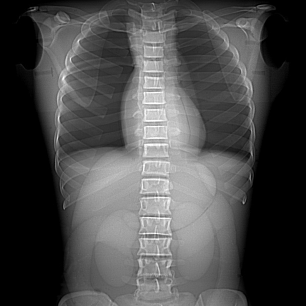
TAKE-APART PIXY is used to demonstrate anatomy as well
as evaluate positioning and imaging techniques,
including kVp, mAs, contrast, optical density, OFD and TFD.
Radiographs of PIXY are optically equivalent to density
and contrast to human patients.
TAKE-APART PIXY allows unlimited exposures and tolerates
trainee errors, permits evaluation of student performance.
- An anatomically and radiologically correct female
- Small size and low weight simplify positioning
- Can be positioned for most views
- Permits evaluation of student performance
- Organs accept contrast media
- Opaque or transparent
Anatomy
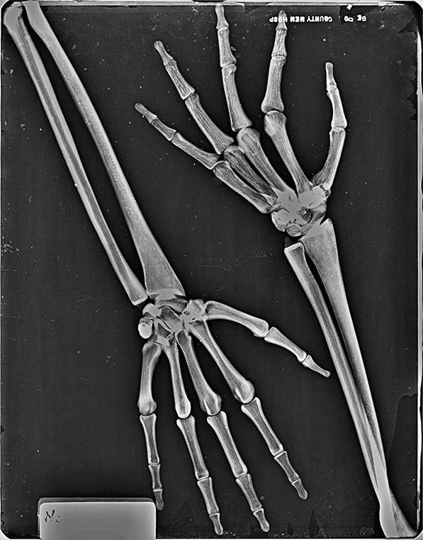
TAKE-APART PIXY’S shoulders have ball and socket joints. Elbows and knees flex 90° to 100°. A “frog position” is possible at the hips. The right hand is molded with fingers positioned for an AP view. The left hand is positioned in oblique-lateral position. The feet are in natural position.
C1, C2, C6, and C7 were converted to mechanical nylon joints because educators in the field prefer full positioning capabilities for the head. This design permits the remaining neck vertebrae to be fixed in a normal position, while assuring a full range of head motion.
TAKE-APART PIXY may be ordered with or without: abdominal and pelvic organs: stomach, gall bladder, urinary bladder, kidneys, rectum, and sigmoid flexure. These are air-filled, but accept water or contrast media and can be easilflushed after use. Custom fractures and custom pathologies are optional at additional cost.
The skull of PIXY has frontal and sphenoidal sinuses, ethmoidal and mastoid air cells and the auditory ossicles. Bone sutures are radiographically visible.
Soft tissues are available in opaque or transparent tissue-equivalent materials. The transparent PIXY has visible organs and skeleton.
Standard PIXY lungs are molded of tissue-equivalent foam with the mass density of inflated human lungs (0.30 g/cc). They are connected to the oro-nasal cavity by the stem bronchi and trachea. The oro-nasal pharynx is filled with a nearly air-equivalent foam.


Delivery Time:
Skeletons are in continuous production. Still, allow
6 – 8 weeks for your PIXY to be made. Production lead time can be
as short as 4 weeks or as long as 12 weeks.
Take-Apart Pixy
Models
Model
- S-103
- RS-103T
- RS-104
- RS-104T
- RS-105
- RS-105T
- RS-157
- RS-102SP
Material
- Opaque
- Transparent
- Opaque
- Transparent
- Opaque
- Transparent
- Actual Lungs (animal) Upgrade
- Custom Fracture or Pathology
Organs & Fill Ports
- Fill Ports & Organs
- Fill Ports & Organs
- Organs Only, NO FILL PORTS
- Organs Only, NO FILL PORTS
- NO ORGANS OR FILL PORTS
- NO ORGANS OR FILL PORTS
- —
- —
RSD In Action
Keele University’s School of Health and Rehabilitation opened a state-of-the-art radiography suite using RSD’s PIXY Phantom to highlight the practice of hands-on training with visual reinforcement.