

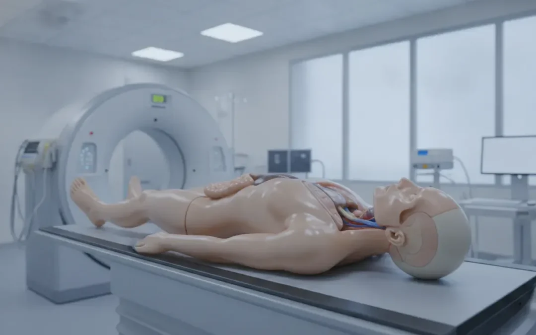
If you are asking what is an anthropomorphic phantom, the short answer is a physical model shaped and constructed to represent the human body, or a body region, during imaging, dosimetry, training, or research. Its anatomy, materials, and internal features are selected to reproduce relevant patient conditions more realistically than a simple plate, cylinder, or uniform block. For the broader definition, see What Is an Imaging Phantom and How Is It Used in Radiology.
Request a quote or custom inquiry from RSD Phantoms to discuss the anatomy, modality, materials, and testing goals your department requires.
What is an anthropomorphic phantom?
An anthropomorphic phantom is a patient-shaped test object that reproduces selected anatomical structures and physical properties of the human body for a defined technical purpose. It may include tissue-equivalent materials, a detailed skeleton, organ inserts, air cavities, motion, dosimetry ports, or imaging markers. The design is not intended to imitate every patient characteristic. It is built to make a specific imaging or measurement question repeatable.
For example, a head phantom may reproduce the skull, cervical spine, and internal landmarks needed for MRI distortion or MRI/CT image-fusion testing. A whole-body model may support positioning practice and repeated radiographic exposures. A sliced radiation therapy phantom may provide locations for dosimeters and materials selected for treatment-planning verification. The word anthropomorphic describes the human-like form and anatomy, while the phantom’s actual capabilities depend on its documented construction.
The National Institute of Standards and Technology describes imaging phantoms as stand-ins for human tissue that help researchers and imaging professionals make controlled, repeatable measurements. That distinction matters: an anthropomorphic phantom is a test and training instrument, not a diagnostic or therapeutic substitute for a patient. NIST’s overview of imaging phantoms provides additional background.
How do anthropomorphic phantoms differ from geometric test objects?
Geometric test objects are often the right choice when a department needs a known, standardized target for one measurement. Anthropomorphic phantoms add anatomical context, which can reveal how positioning, tissue interfaces, organ locations, bone structure, and field of view affect an imaging workflow.
| Design type | What it represents | Typical questions it helps answer |
|---|---|---|
| Geometric test object | Known shapes, densities, rods, spheres, or step patterns | Is uniformity, contrast, resolution, dose, or calibration within the expected range? |
| Anthropomorphic phantom | Human-like anatomy with selected tissue and organ features | How does the system or protocol perform with realistic anatomy, positioning, attenuation, or organ relationships? |
| Hybrid or multimodality model | Anatomy plus features for more than one imaging method | Can teams compare, register, or align images across modalities using the same physical anatomy? |
A department may use both types. A geometric QA device can provide a fast, repeatable check of a specific parameter, while an anthropomorphic model can support a broader workflow assessment. The choice should follow the measurement objective, the modality, the required level of anatomical realism, and the department’s operating procedure.
Which materials make an anthropomorphic phantom realistic?
The word realistic should be tied to the property being tested. A phantom can be anatomically detailed but unsuitable for a particular modality if its materials do not reproduce the relevant attenuation, density, relaxation, acoustic, or radioactive behavior. Good phantom design therefore combines anatomy with materials that have been selected and characterized for the intended application.
Tissue-equivalent materials
Tissue-equivalent materials are formulated to approximate one or more physical properties of human tissues. Depending on the application, a design may include substitutes for soft tissue, lung, adipose tissue, breast tissue, cortical bone, trabecular bone, liver, kidney, or other structures. The target property may be x-ray attenuation, CT number, electron density, MRI signal behavior, ultrasound response, or another measurable characteristic.
Material equivalence is never automatically universal. A material that performs well for an x-ray or CT task does not necessarily reproduce the MRI, ultrasound, or radiation-therapy behavior needed for a different test. A useful specification should identify the modality, energy range, tissue property, and measurement method it supports. The peer-reviewed review Tissue mimicking materials for imaging and therapy phantoms surveys how material requirements vary across imaging and therapy applications.
Real cadaver-bone options
Some anthropomorphic phantoms are available with authentic human cadaver bones. This option can add real skeletal anatomy and material variation to applications where bone structure and radiographic detail matter. It is a specialized configuration, not a feature that should be assumed for every phantom or every model.
RSD Phantoms documents cadaver-bone configurations for products such as the Take-Apart Pixy with Cadaver Bones, the ART Phantom with Cadaver Bones, and selected head phantom designs. A department considering this option should ask about the specific model, included anatomy, documentation, handling requirements, and the imaging or training objective. The Take-Apart Pixy with Cadaver Bones page describes one product-specific configuration with a nine-part modular body and optional organs.
Organ inserts, cavities, and measurement features
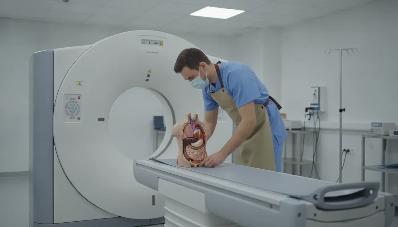
Organ inserts and internal cavities let a phantom represent more than an outer silhouette. Depending on the design, organs may be removable, fillable, replaceable, or configured for a particular anatomy. Air spaces can be important in chest and head imaging. Markers, dosimetry holes, rods, spheres, and motion components can create measurable targets inside the anatomical model.
These features should be treated as configuration decisions. A buyer should confirm which organs are included, whether they accept water or contrast media, how they are positioned, and whether replacement or custom components are available. For example, RSD’s Take-Apart Pixy configurations can include abdominal and pelvic organs, while the Multi-Modality Fusion Head Phantom is designed around MRI/CT registration and distortion evaluation rather than whole-body organ training.
Where are anthropomorphic phantoms used?
Anthropomorphic phantoms are used when a team needs a controlled physical model that preserves meaningful anatomical context. The same broad design principle can support different work across departments, but each use case requires its own performance criteria and documented setup.
- Radiography and CT: Practice positioning, evaluate technique choices, assess image quality, and compare repeatable protocols with a patient-shaped model.
- MRI: Assess geometric distortion, image registration, coil fit, and multimodality alignment when the design supports those tests.
- Radiation therapy: Support dosimetry, treatment-planning checks, image guidance, and end-to-end QA when the phantom includes suitable tissues, interfaces, and detector locations.
- Nuclear medicine: Evaluate organ-specific or image-quality workflows when the phantom is designed for the relevant radiotracer, geometry, and measurement method.
- Education and skills training: Give radiologic technologists and students a repeatable subject for anatomy recognition, positioning, exposure practice, and feedback without relying on repeated patient participation.
- Research and development: Provide a controlled platform for comparing protocols, reconstruction methods, devices, software, or image-guided workflows.
RSD’s portfolio spans medical imaging, quality assurance, multi-modality imaging, radiation therapy, nuclear medicine, and health physics. Explore the medical imaging phantom category for training and anatomical imaging applications, or review multi-modality phantoms when the project involves MRI, CT, and X-ray relationships.
Contact RSD Phantoms for a modality-specific recommendation before selecting a configuration. A technical conversation can clarify which anatomy, materials, inserts, and documentation fit the intended test.
How should a department select an anthropomorphic phantom?
The best anthropomorphic phantom is defined by the question your department needs to answer, not by appearance alone. Use the following sequence to turn a broad request into a defensible specification.
- Define the primary job: Decide whether the phantom is for education, positioning, routine QA, acceptance testing, protocol development, dosimetry, image registration, research, or a combination.
- Name the modality and operating range: Identify the imaging systems, energy or acquisition conditions, field of view, coils, detectors, or radiotracer requirements involved.
- Specify the anatomy: Choose the body region, sex or body habitus representation, organ configuration, skeletal detail, pathologies, trauma features, motion, and disassembly needs.
- Match materials to the measurement: Ask which tissue properties are represented, how they were characterized, and whether the documentation supports the intended modality and energy range.
- List measurement interfaces: Confirm the need for dosimetry holes, fill ports, removable inserts, markers, rods, spheres, reference landmarks, or software-compatible targets.
- Plan repeatability and handling: Consider setup time, storage, transport, cleaning, replacement parts, training, and whether the model must withstand frequent student use.
- Request documentation and customization details: Ask for a technical sheet, supported applications, configuration options, validation information, and a quote based on the final specification.
For products with documented tissue-equivalent or standards alignment, review the product-specific documentation rather than applying the claim to an entire catalog. For example, RSD documents ICRU-44 alignment for its ART Phantom line. That statement should not be generalized to every RSD product without supporting documentation.
Frequently Asked Questions
What is an anthropomorphic phantom used for?
An anthropomorphic phantom is used to represent human anatomy during imaging, dosimetry, education, quality assurance, protocol development, or research. Its exact use depends on the documented anatomy, materials, inserts, motion, and measurement interfaces. It is a controlled test or training instrument, not a replacement for clinical patient care.
Are anthropomorphic phantoms made from tissue-equivalent materials?
Many anthropomorphic phantoms use one or more tissue-equivalent materials, but the material properties are product- and modality-specific. A buyer should confirm which tissues are represented, which physical properties are matched, and the energy or acquisition range covered by the documentation before selecting a phantom for a particular test.
Why would a phantom include real cadaver bones?
Real cadaver bones can provide authentic skeletal anatomy and material variation for applications where bone detail matters. They are a specialized option available in selected configurations, not a universal feature. Departments should confirm sourcing documentation, included anatomy, handling requirements, and the intended imaging or training use with the manufacturer.
What is the difference between an anthropomorphic phantom and a QA phantom?
Anthropomorphic describes a human-like anatomical form, while QA describes a purpose: checking system or workflow performance against a known expectation. An anthropomorphic phantom can support QA, but many QA phantoms are geometric plates, cylinders, or modular test objects. The right choice depends on the measurements and anatomical context required.
Can one anthropomorphic phantom be used for every imaging modality?
No. Some designs support multiple modalities, but no single configuration should be assumed to reproduce every relevant tissue property or workflow. Confirm the manufacturer’s documented modality support, materials, field of view, markers, inserts, and measurement method for each planned application.
Request a quote from Radiology Support Devices Inc. to discuss an anthropomorphic phantom for medical imaging, education, quality assurance, radiation therapy, nuclear medicine, health physics, or research.