Multi-Modality Fusion Head Phantom
VALIDATION OF MRI/CT FUSION IMAGES USING GEOMETRIC DISTORTION MEASUREMENTS
- Anthropomorphic head phantom for MRI and CT imaging
- Evaluate MRI image distortion in Stereotactic Radiosurgery (SRS) Planning
- Versatile verification phantom for image fusion and deformable image registration software across treatment planning systems
- Ideal for MRI sequence optimization
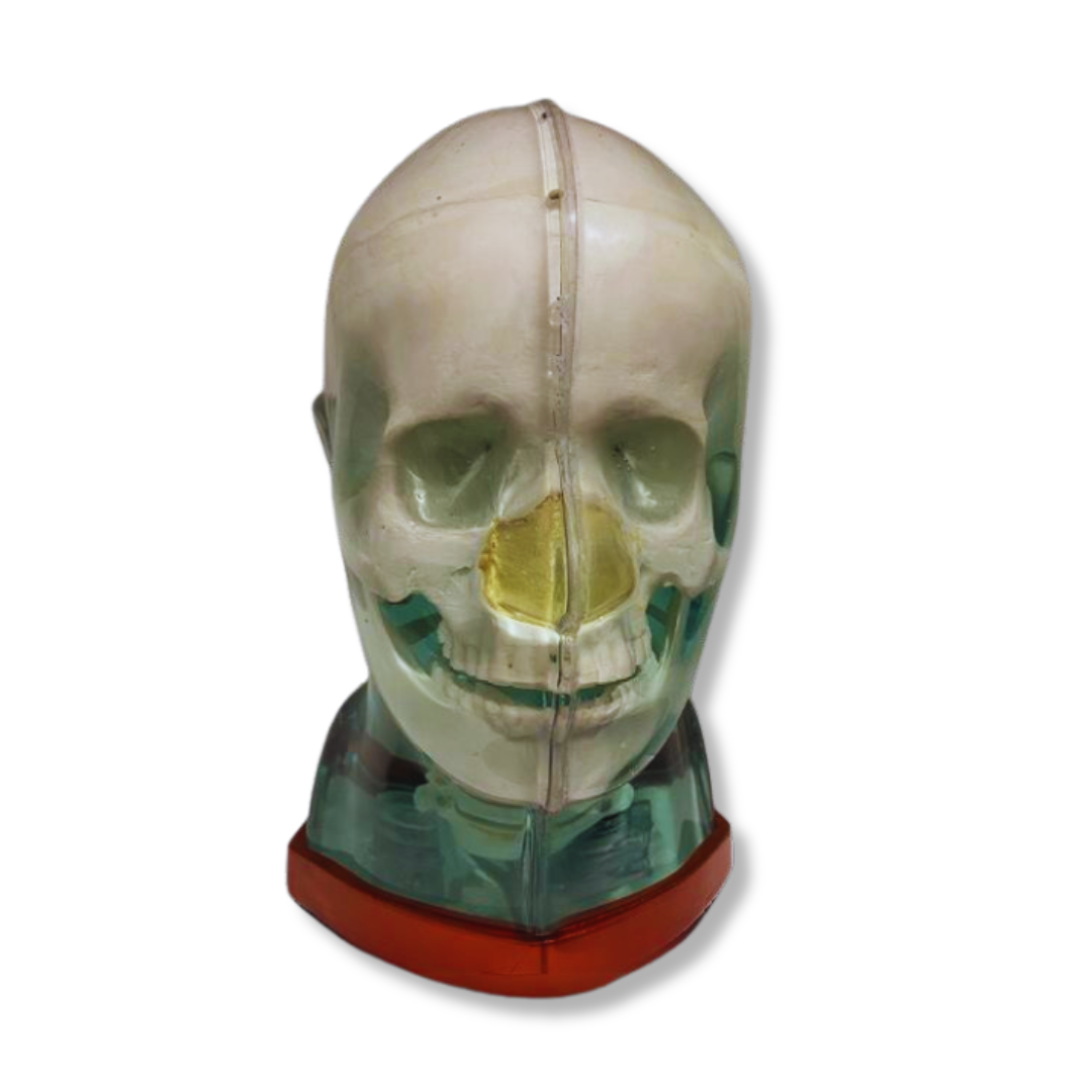
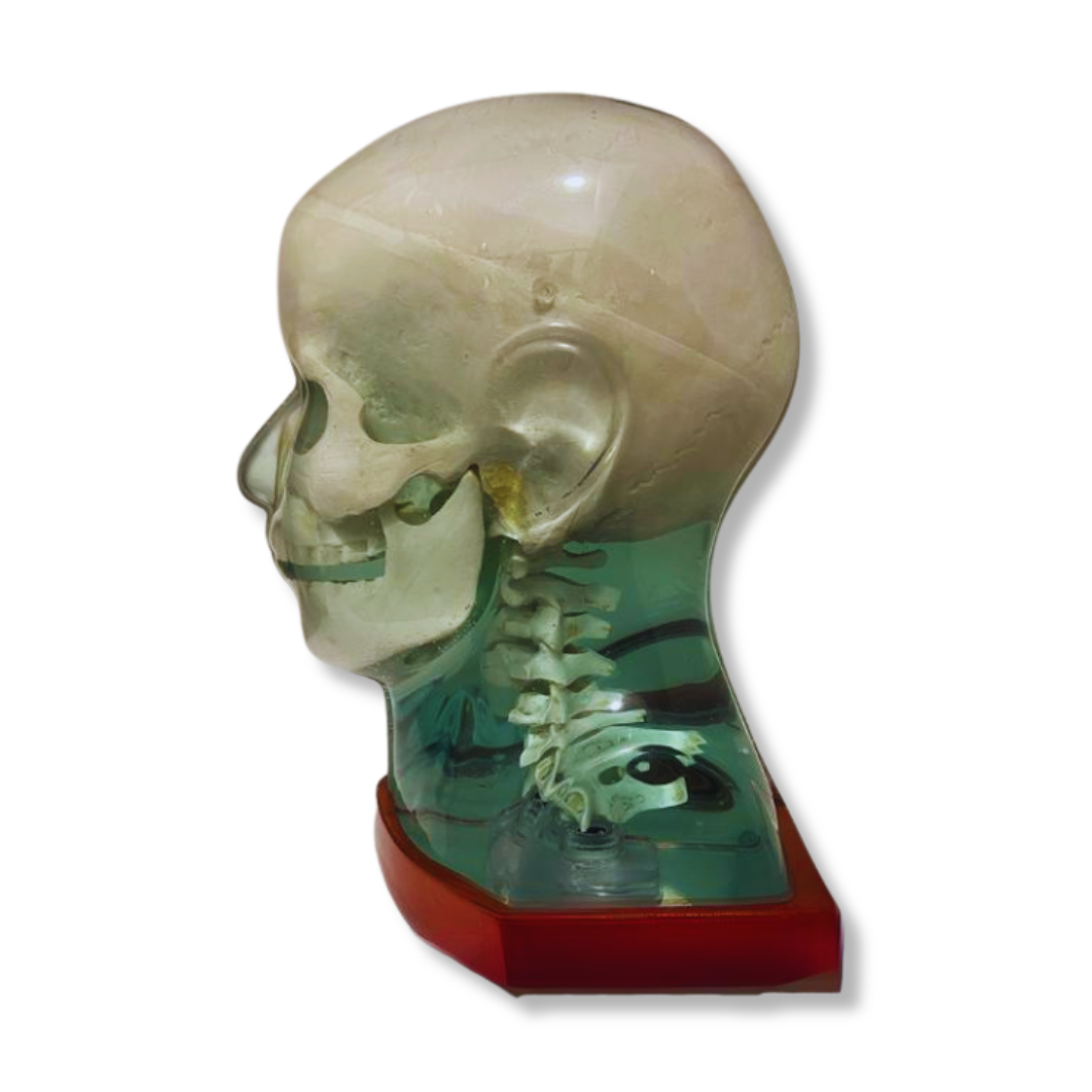
Experience Precision Imaging Assessment with RSD’s Multi-Modality Fusion Head Phantom, offering comprehensive evaluations across MRI, CT, and X-ray modalities. This cutting-edge anthropomorphic phantom showcases an intricately detailed skull and cervical spine, faithfully replicating the attenuation properties of both trabecular and cortical bone.
Jump To: Accessories | Model Number | Applications & Modalities | Clinical Images
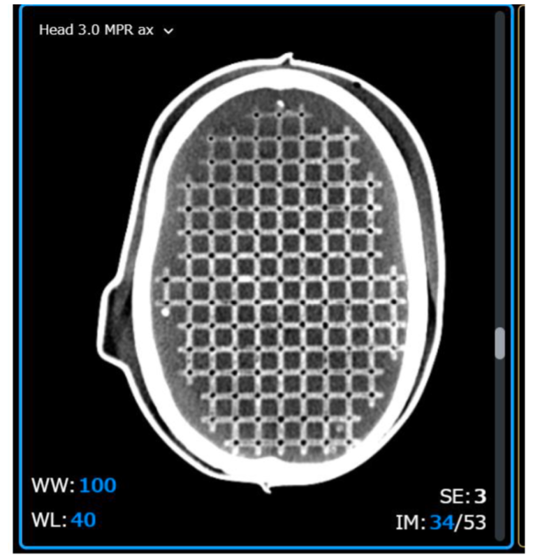
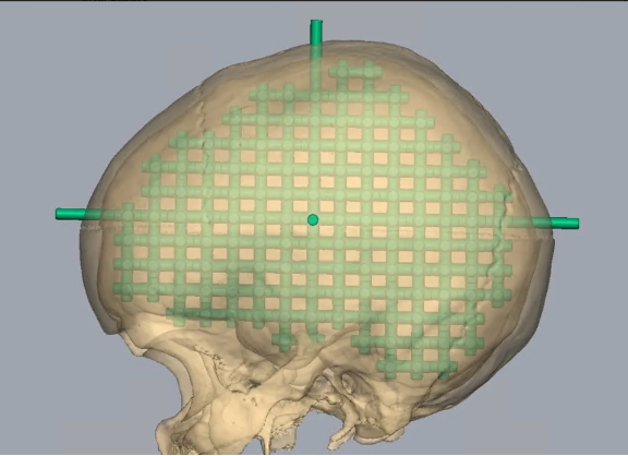
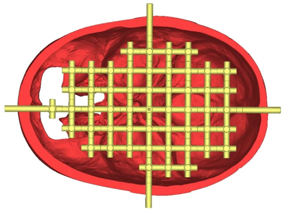
For lifelike MRI and CT imaging, our proprietary aqueous solution generates soft tissue signals, allowing for T1, T2, and PD weighted imaging. Within the cranial section, a dedicated 3D lattice is meticulously designed to evaluate MR distortion within the head scan field of view.
To ensure structural integrity and contain the aqueous solution securely, the entire phantom is encased in a transparent, scatter-resistant plastic shell.
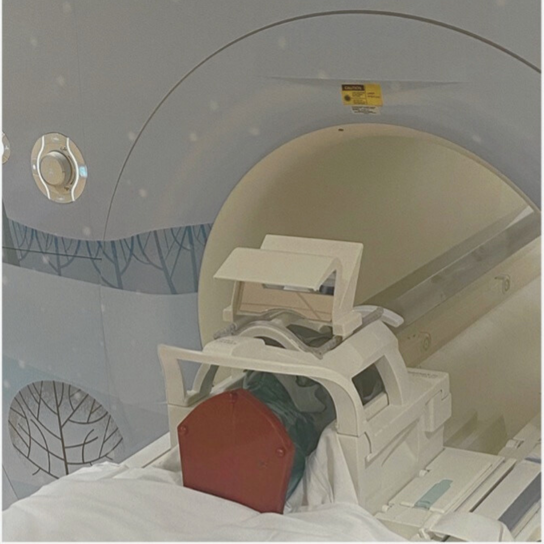
Designed to fit perfectly in the Siemens and GE head coil, our Multi-Modality Fusion Head Phantom serves as a reliable quality assurance tool for both CT and MRI applications. Additionally, it proves to be an indispensable for protocol optimization and technologist training.
In MRI, technicians can assess magnetic field distortion by utilizing the 3D printed brain lattice within the cranial section. This lattice, filling the entire intracranial space, is composed of 2.5 mm diameter circular rods spaced at 10 mm intervals (I-S), 10.5 mm intervals (AP), and 11 mm intervals (L-R). At the lattice’s center, 5mm rods establish the x-y-z axes, aiding users in orienting the lattice within the field of view. The phantom includes MRI/CT compatible markers at the ends of these rods, facilitating co-registration alignment in fused images.
For CT applications, technicians can evaluate image quality and geometric distortion through the 3D lattice located within the cranium. This phantom offers a meticulously detailed skull and cervical spine crafted from materials simulating trabecular and cortical bone, accurately mimicking the attenuation properties of a human skull. This fidelity holds true across diagnostic and therapeutic energy ranges, spanning from 50 keV to 25 MeV.
Accessories
- Comprehensive Product Manual
Model Number
| RS-MMH | Multi-Modality Head Phantom |
Applications
- MRI/CT fusion image verification
- MRI geometric distortion measurements in small FOV for SRS & diagnostic imaging
- Teaching & training
- Image quality
- Protocol verification
- Applicable MRI sequences: T1 FSE, T2 FSE, MPRAGE and CISS, Proton Density
Modalities
- MRI
- CT
- Cone beam CT
- X-ray
- Panoramic X-ray
- Fluoroscopy
- PET
- SPECT
Clinical Images