


Take-Apart Pixy
IDEAL SUBSTITUTE FOR TEACHING & TRAINING RADIOLOGICAL TECHNOLOGISTS
- NEW! Inclusive skin tones, from light to dark complexion, available at no additional cost
- Anthropomorphic phantom disassembles into 9 parts
- Small size and low weight simplify positioning
- Unlimited repetition of most views for which patients cannot be used
- Provides valid feedback to evaluate trainee performance
- Designed to image any clinical view (AP, oblique, lateral, frog legs, etc.)
- Custom pathologies and traumas available
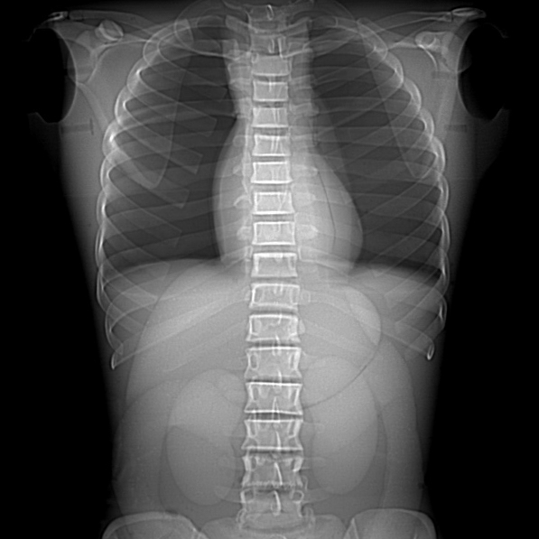
Offered in opaque or transparent, Take-Apart Pixy is an anatomically and radiologically correct female designed specifically for training radiologic technologists. At 5’ 1” (156 cm) weighing 105 lbs (48 kg), Take-Apart Pixy is a repeatable, convenient substitute for patients and virtually indestructible.
Take-Apart Pixy may be ordered with or without abdominal and pelvic organs: stomach, gall bladder, urinary bladder, kidneys, rectum, and sigmoid flexure. The organs are air-filled but accept water or contrast media that can be easily flushed after use. Custom pathologies and traumas are available at an additional cost.
Built with soft-tissue mold and skeleton molds that are matched for anatomic fidelity, Take-Apart Pixy permits unlimited exposures, demonstrates the effects of changing technical factors, and allows for the evaluation of student performance. Students have no difficulty in maneuvering Take-Apart Pixy into the most desired positions as the phantom is built to tolerate trainee errors.
Take-Apart Pixy is used to demonstrate anatomy and evaluate positioning and imaging techniques, including kVp, mAs, contrast, optical density, digital processing, OFD, and TFD. Made of tissue-equivalent materials and life-like articulations, Take-Apart Pixy is more realistic than a cadaveric skeleton with radiographs that are optically equivalent in density and contrast to human patients.
C1, C2, and C7 vertebrae were converted to mechanical nylon joints because educators in the field prefer full positioning capabilities for the head. This design permits the remaining neck vertebrae to be fixed in a normal position while assuring a full range of head motion.
The skull of Take-Apart Pixy has frontal and sphenoidal sinuses, ethmoidal and mastoid air cells, and the auditory ossicles. Bone sutures are radiographically visible.
Soft tissues are available in opaque or transparent tissue-equivalent materials. The transparent Take-Apart Pixy has visible organs and skeleton.
Take-Apart Pixy lungs are molded of tissue-equivalent foam with the mass density of inflated human lungs (0.30 g/cc). They are connected to the oro-nasal cavity by the stem bronchi and trachea. The oro-nasal pharynx is filled with a nearly air-equivalent foam.
Model Numbers
| RS-103 | Opaque | Take-Apart Pixy with Fill Ports and Organs. Contains fill ports for the stomach, gall bladder, urinary bladder, right and left kidneys, rectum, and sigmoid flexor. Includes storage case. |
| RS-103T | Transparent | Take-Apart Pixy with Fill Ports and Organs. Contains fill ports for the stomach, gall bladder, urinary bladder, right and left kidneys, rectum, and sigmoid flexor. Includes storage case. |
| RS-104 | Opaque | Take-Apart Pixy with Organs but NO Fill Ports. Contains stomach, gall bladder, urinary bladder, right and left kidneys, rectum, and sigmoid flexor. Includes storage case. |
| RS-104T | Transparent | Take-Apart Pixy with Organs but NO Fill Ports. Contains stomach, gall bladder, urinary bladder, right and left kidneys, rectum, and sigmoid flexor. Includes storage case. |
| RS-105 | Opaque | Take-Apart Pixy with NO Fill Ports and NO Organs. Includes storage case. |
| RS-105T | Transparent | Take-Apart Pixy with NO Fill Ports and NO Organs. Includes storage case. |
Please contact RSD for custom pathologies, traumas, and inclusive skin tone options.
Applications
- Teaching & training of patient positioning
- Image quality
- Diagnostic radiology
- Dosimetry verification
- Protocol verification
Modalities
- CT
- X-Ray
- Fluoroscopy
Anatomy
- Shoulders have ball and socket joints
- Elbows and knees flex 90° to 100°
- A “frog position” is possible at the hips
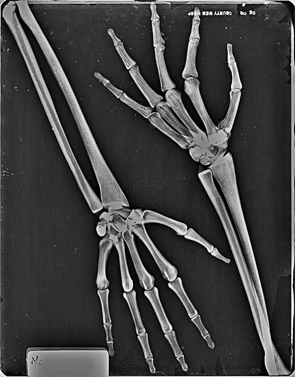
- Right hand is molded with fingers positioned for an AP view
- Left hand is in an oblique-lateral position
- Feet are in natural position
Clinical Images
Publication References
Xu Q, Tong X, Lin M, et al. Time and Frequency to Observe Fiducial Markers in MLC-Modulated Fields During Prostate IMRT/VMAT Beam Delivery. Phys Med. 2020;76:142-149. DOI: 10.1016/j.ejmp.2020.06.026
Paysan P. et al. (2020) Deep Learning Methods for Image Guidance in Radiation Therapy. In: Schilling FP., Stadelmann T. (eds) Artificial Neural Networks in Pattern Recognition. ANNPR 2020. Lecture Notes in Computer Science, vol 12294. Springer, Cham. DOI: 10.1007/978-3-030-58309-5_1
Holmström A. Radiography Students’ Learning of Plain X-Ray Examinations in Simulation Laboratory Exercises: An Ethnographic Research. Journal of Medical Imaging and Radiation Sciences. 2019 Dec. DOI: 10.1016/j.jmir.2019.07.005
RSD In Action
Keele University’s School of Health and Rehabilitation opened a state-of-the-art radiography suite using RSD’s PIXY Phantom to highlight the practice of hands-on training with visual reinforcement.